logo
AAT Bioquest

Rhod-4™, AM

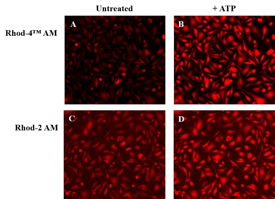
Calcium measurement is critical for numerous biological investigations. Fluorescent probes that show spectral responses upon binding Ca2+ have enabled researchers to investigate changes in intracellular free Ca2+ concentrations by using fluorescence microscopy, flow cytometry, fluorescence spectroscopy and fluorescence microplate readers. Rhod-2 is most commonly used among the red fluorescent calcium indicators. However, Rhod-2 AM is only moderately fluorescent in live cells upon esterase hydrolysis, and has very small cellular calcium responses. Rhod-4™ has been developed to improve Rhod-2 cell loading and calcium response while maintaining the spectral wavelength of Rhod-2. In CHO and HEK cells Rhod-4™ AM has cellular calcium response that is 10 times more sensitive than Rhod-2 AM. AAT Bioquest offers versatile packing sizes of Quest Rhod-4 to meet your special needs, e.g., 1 mg; 10x50 µg; 20x50 µg; HTS packages with no additional packaging charge.
Example protocol

PREPARATION OF STOCK SOLUTIONS

Unless otherwise noted, all unused stock solutions should be divided into single-use aliquots and stored at -20 °C after preparation. Avoid repeated freeze-thaw cycles

Rhod-4™ AM Stock Solution
  1. Prepare a 2 to 5 mM stock solution of Rhod-4™ AM in high-quality, anhydrous DMSO.

PREPARATION OF WORKING SOLUTION

Rhod-4™ AM Working Solution
  1. On the day of the experiment, either dissolve Rhod-4™ AM in DMSO or thaw an aliquot of the indicator stock solution to room temperature.

  2. Prepare a 2 to 20 µM Rhod-4™ AM working solution in a buffer of your choice (e.g., Hanks and Hepes buffer) with 0.04% Pluronic® F-127. For most cell lines, Rhod-4™ AM at a final concentration of 4-5 μM is recommended. The exact concentration of indicators required for cell loading must be determined empirically.

    Note: The nonionic detergent Pluronic® F-127 is sometimes used to increase the aqueous solubility of Rhod-4™ AM. A variety of Pluronic® F-127 solutions can be purchased from AAT Bioquest.

    Note: If your cells contain organic anion-transporters, probenecid (1-2 mM) may be added to the dye working solution (final in well concentration will be 0.5-1 mM) to reduce leakage of the de-esterified indicators. A variety of ReadiUse™ Probenecid products, including water-soluble, sodium salt, and stabilized solutions, can be purchased from AAT Bioquest.

SAMPLE EXPERIMENTAL PROTOCOL

Following is our recommended protocol for loading AM esters into live cells. This protocol only provides a guideline and should be modified according to your specific needs.

  1. Prepare cells in growth medium overnight.
  2. On the next day, add 1X Rhod-4™ AM working solution to your cell plate.

    Note: If your compound(s) interfere with the serum, replace the growth medium with fresh HHBS buffer before dye-loading.

  3. Incubate the dye-loaded plate in a cell incubator at 37 °C for 30 to 60 minutes.

    Note: Incubating the dye for longer than 1 hour can improve signal intensities in certain cell lines.

  4. Replace the dye working solution with HHBS or buffer of your choice (containing an anion transporter inhibitor, such as 1 mM probenecid, if applicable) to remove any excess probes.
  5. Add the stimulant as desired and simultaneously measure fluorescence using either a fluorescence microscope equipped with a TRITC filter set or a fluorescence plate reader containing a programmable liquid handling system such as an FDSS, FLIPR, or FlexStation, at Ex/Em = 540/590 nm cutoff 570 nm.

Calculators

Common stock solution preparation

Table 1. Volume of DMSO needed to reconstitute specific mass of Rhod-4™, AM to given concentration. Note that volume is only for preparing stock solution. Refer to sample experimental protocol for appropriate experimental/physiological buffers.

0.1 mg0.5 mg1 mg5 mg10 mg
1 mM98.429 µL492.145 µL984.291 µL4.921 mL9.843 mL
5 mM19.686 µL98.429 µL196.858 µL984.291 µL1.969 mL
10 mM9.843 µL49.215 µL98.429 µL492.145 µL984.291 µL

Molarity calculator

Enter any two values (mass, volume, concentration) to calculate the third.

Mass (Calculate)Molecular weightVolume (Calculate)Concentration (Calculate)Moles
/=x=

Spectrum

Product family

Citations

View all 59 citations: Citation Explorer
Brain Slice Derived Nerve Fibers Grow along Microcontact Prints and are Stimulated by Beta-Amyloid (42)
Authors: Steiner, Katharina and Humpel, Christian
Journal: Frontiers in Bioscience-Landmark (2024): 232
Simultaneous Widefield Voltage and Dye-Free Optical Mapping Quantifies Electromechanical Waves in Human Induced Pluripotent Stem Cell-Derived Cardiomyocytes
Authors: Liu, Wei and Han, Julie L and Tomek, Jakub and Bub, Gil and Entcheva, Emilia
Journal: ACS Photonics (2023)
High-content analysis and Kinetic Image Cytometry identify toxicity and epigenetic effects of HIV antiretrovirals on human iPSC-neurons and primary neural precursor cells
Authors: Smith, Alyson S and Ankam, Soneela and Farhy, Chen and Fiengo, Lorenzo and Basa, Ranor CB and Gordon, Kara L and Martin, Charles T and Terskikh, Alexey V and Jordan-Sciutto, Kelly L and Price, Jeffrey H and others,
Journal: Journal of Pharmacological and Toxicological Methods (2022): 107157
RGS5 Attenuates Baseline Activity of ERK1/2 and Promotes Growth Arrest of Vascular Smooth Muscle Cells
Authors: Demirel, Eda and Arnold, Caroline and Garg, Jaspal and J{\"a}ger, Marius Andreas and Sticht, Carsten and Li, Rui and Kuk, Hanna and Wettschureck, Nina and Hecker, Markus and Korff, Thomas
Journal: Cells (2021): 1748

References

View all 34 references: Citation Explorer
Fluorescence absorbance inner-filter decomposition: the role of emission shape on estimates of free Ca(2+) using Rhod-2
Authors: Territo PR, Heil J, Bose S, Evans FJ, Balaban RS.
Journal: Appl Spectrosc (2007): 138
Protein kinase C and myocardial calcium handling during ischemia and reperfusion: lessons learned using Rhod-2 spectrofluorometry
Authors: Stamm C, del Nido PJ.
Journal: Thorac Cardiovasc Surg (2004): 127
Novel fluo-4 analogs for fluorescent calcium measurements
Authors: Martin VV, Beierlein M, Morgan JL, Rothe A, Gee KR.
Journal: Cell Calcium (2004): 509
Kinetic characterization of novel NR2B antagonists using fluorescence detection of calcium flux
Authors: Bednar B, Cunningham ME, Kiss L, Cheng G, McCauley JA, Liverton NJ, Koblan KS.
Journal: J Neurosci Methods (2004): 247
Cytosolic calcium in the ischemic rabbit heart: assessment by pH- and temperature-adjusted rhod-2 spectrofluorometry
Authors: Stamm C, Friehs I, Choi YH, Zurakowski D, McGowan FX, del Nido PJ.
Journal: Cardiovasc Res (2003): 695
Page updated on November 21, 2024

Ordering information

Price
AvailabilityIn stock
Unit size
1 mg
5x50 ug
10x50 ug
20x50 ug
Catalog Number
21120211212112221123
Quantity
Add to cart

Additional ordering information

Telephone1-800-990-8053
Fax1-800-609-2943
Emailsales@aatbio.com
InternationalSee distributors
Bulk requestInquire
Custom sizeInquire
Technical SupportContact us
Purchase orderSend to sales@aatbio.com
ShippingStandard overnight for United States, inquire for international
Request quotation

Physical properties

Dissociation constant (Kd, nM)451

Molecular weight

1015.96

Solvent

DMSO

Spectral properties

Excitation (nm)

523

Emission (nm)

551

Quantum yield

0.11

Storage, safety and handling

Certificate of OriginDownload PDF
H-phraseH303, H313, H333
Hazard symbolXN
Intended useResearch Use Only (RUO)
R-phraseR20, R21, R22

Storage

Freeze (< -15 °C); Minimize light exposure
UNSPSC12352200

Platform

Fluorescence microscope

ExcitationTRITC filter set
EmissionTRITC filter set
Recommended plateBlack wall, clear bottom

Fluorescence microplate reader

Excitation540
Emission590
Cutoff570
Recommended plateBlack wall, clear bottom
Instrument specification(s)Bottom read mode, Programmable liquid handling
ATP-stimulated calcium responses of endogenous P2Y receptors were measured in CHO-K1 cells with Rhod-4&trade; AM (Cat# 21120) and Rhod-2 AM (Cat# 21064). CHO-K1 cells were seeded overnight at 50,000 cells/100 &micro;L/well in a Costar 96-well black wall/clear bottom plate. The growth medium was removed, and the cells were incubated with 100 &micro;L of dye loading solution using Rhod-4&trade; AM (4 &micro;M, A and B) or Rhod-2 AM (4 &micro;M, C and D) for 1 hour in a 37 &deg;C, 5% CO2 incubator. The staining solution was replaced with 200 &micro;L HHBS, then the cells were imaged before (A and C) and after (B and D) ATP treatment with a fluorescence microscope (Olympus IX71) using TRITC channel.
ATR effects on cardiomyocyte electrophysiology. (A) Action potentials in response to electrical pacing were imaged optically (using di4-ANBDQBS) and presented as mean&thinsp;&plusmn;&thinsp;SEM at each point for each group. (B) Quantified APD80 for control CM and ChR2-CM without ATR and with 1&thinsp;&mu;M ATR (highest optical excitability). Both A and B had n&thinsp;=&thinsp;3&ndash;4 samples per experimental group. (C) Calcium transients in response to electrical pacing were imaged optically (using Rhod4-AM) and presented as mean&thinsp;&plusmn;&thinsp;SEM at each point for each group. (D) Quantified CTD80 for control CM and ChR2-CM at different ATR supplements. Both C and D had n&thinsp;=&thinsp;7&ndash;33 samples for each of the eight experimental groups. (E) Example activation maps of ChR2-CM, following point electrical stimulation at the bottom; isochrones are 10&thinsp;ms apart. Scale bar is 5&thinsp;mm. (F) Quantified conduction velocity from the activation maps (n&thinsp;=&thinsp;7&ndash;14 per group). (*) indicates significant difference at p&thinsp;&lt;&thinsp;0.05 compared to the respective control (zero ATR) or as indicated by the brackets. Source: <strong>Cardiac Optogenetics: Enhancement by All-trans-Retinal </strong>by Yu et al., <em>Scientific Reports</em>, Nov. 2016.
GPR157 couples with Gq-class of the heterotrimeric G-proteins. (A&ndash;C) Plasmids expressing indicated protein were transfected into U-2 OS cells. Rhod-4, a fluorescent calcium indicator, were used to assess changes in [Ca<sup>2+</sup>]i. Application of Ionomycin, a calcium ionopohore, to cells after experiments confirmed almost uniform uptake of Rhod-4 in these cells. (D&ndash;F) Fluorescent intensity of Rhod4 in GFP-positive cells. Mean&thinsp;&plusmn;&thinsp;s.e.m. Data were obtained from 3 independent experiments (more than 40 cells). **p&thinsp;&lt;&thinsp;0.01, ***p&thinsp;&lt;&thinsp;0.001. Scale bar: 10&thinsp;&mu;m. Source: <strong>The G protein-coupled receptor GPR157 regulates neuronal differentiation of radial glial progenitors through the Gq-IP3 pathway </strong>by Takeo et al., <em>Scientific Reports</em>, May 2016.
Nuclear calcium transients in neonatal rat ventricular cardiomyocytes (NRVCs) exposed to hypertrophic stimuli. A. Original recording of only cytoplasmic Ca<sup>2+</sup> transients in NRVC with fluo-4. B. Line scan imaging of only cytoplasmic Ca<sup>2+</sup> transients in cardiomyocytes with fluo-4. C. Original recording of only nucleus Ca<sup>2+</sup> transients in cardiomyocytes with fluo-4. D. Line scan imaging of cytoplasmic Ca<sup>2+</sup> transients in cardiomyocytes with fluo-4. E. Original recording of cytoplasmic and nucleus Ca<sup>2+</sup> transients in NRVC with a different Ca indicator, Rhod-4. F. Line scan imaging of only cytoplasmic Ca<sup>2+</sup> transients in cardiomyocytes with Rhod-4. G. Line scan imaging of nuclear Ca<sup>2+</sup> transients in cardiomyocytes with Rhod-4. H. Average values of the effects of hypertrophic stimuli on time to F/F0 in the cytoplasm of NRVCs exposed to vehicle (control; n = 19), Ang II (n = 42), ET-1 (n = 21), or PE (n = 29). I. Average values of the effects by hypertrophic stimuli on time to F/F0 in the nucleus of NRVCs exposed to vehicle (control; n = 19), Ang II (n = 42), ET-1 (n = 21), or PE (n = 29). *P &lt; 0.05 compared to the control. **P &lt; 0.01 compared to the control. &dagger;P &lt; 0.001 compared to the control.&nbsp;Source: <strong>Emerin plays a crucial role in nuclear invagination and in the nuclear calcium transient</strong> by Shimojima et al., <em>Scientific Reports,</em> March 2017.
Response to optical stimulation in light-sensitive cardiac syncytia. (a,b) Activation maps resulting from optical stimulation (1&thinsp;Hz) of <em>in vitro</em> light-sensitive cell monolayers in the island configuration. Optical stimulus strength was at most 0.07&thinsp;mW/mm<sup>2</sup> greater than the threshold irradiance required to elicit a propagating response (E<sub>e,thr</sub>). Time zero corresponds to the beginning of a 20&thinsp;ms-long pulse of blue light (wavelength &lambda;&thinsp;=&thinsp;470&thinsp;nm) applied to the 1&thinsp;cm-diameter region indicated by the dashed black line in (a); spacing between isochrones is 10&thinsp;ms. (c,d) Same as (a,b) but for <em>in silico</em> cell monolayers. Simulated optical stimuli were at most 0.0005&thinsp;mW/mm<sup>2</sup> greater than E<sub>e,thr</sub>. Here time zero corresponds to the end of each 20&thinsp;ms-long illumination pulse instead of the beginning; spacing between isochrones is 10&thinsp;ms. Black-coloured locations did not activate. (e,f) Select <em>in vitro </em>calcium transients from the pixel locations 1&ndash;4 indicated in (a,b) on opposite sides of the island of ChR2-expressing donor cells (CM in GD and HEK in CD) showing the wavefront activation sequence. (g,h) Select <em>in silico</em> voltage traces (analogous to those in (e,f)) from locations 1&ndash;4. Source:<strong> Optogenetics-enabled assessment of viral gene and cell therapy for restoration of cardiac excitability</strong> by Ambrosi et al., <em>Scientific Reports</em>, Dec. 2015.