logo
AAT Bioquest

MitoDNA™ Red 680

Product key features

  • mtDNA-Specific: Selectively binds to mtDNA without nuclear interference
  • Live-Cell Compatibility: Easily permeates cells for real-time mtDNA tracking
  • High Signal Clarity: Large Stokes shift ensures a strong signal-to-noise ratio for multiplex imaging
  • Broad Research Applicability: Ideal for investigating mtDNA-linked diseases and mitochondrial function
Product description

There are limited probes available that effectively detect mitochondrial deoxyribonucleic acid (mtDNA). Common fluorescent DNA probes, such as DAPI, Hoechst, or SYBR® Green, lack the specificity required for mitochondrial targeting, primarily staining nuclear DNA. MitoDNA™ Red 680 is a cell-permeable dye that specifically stains mtDNA in live cells, offering an efficient method for the dynamic imaging of mtDNA. This dye exhibits a large Stokes shift, providing an excellent signal-to-noise ratio and enabling easy multiplex staining with other fluorescent probes. mtDNA is a small, circular DNA molecule located within mitochondria in the cytoplasm. It is supplementary to nuclear DNA and encodes 37 genes essential for mitochondrial and cellular functions. Mitochondria are responsible for ATP synthesis through oxidative phosphorylation and contain the genetic information for synthesizing key enzymes, transfer RNA (tRNA), and ribosomal RNA (rRNA). Mutations and disorders in mtDNA can lead to a range of health issues, including age-related hearing loss, diabetes, and failures in the brain, heart, and liver. Additionally, mtDNA mutations are associated with an increased risk of various cancers, including lymphomas, leukemias, and tumors in the breast, intestines, liver, and kidneys.

Example protocol

AT A GLANCE

Important Note

Before using MitoDNA™ Red 680 for the first time, allow it to thaw at room temperature. Then, briefly centrifuge it to collect the dried pellet.

Protocol Summary
  1. Prepare cells in a growth medium.

  2. Stain cells with MitoDNA™ Red 680 working solution.

  3. Incubate samples for 5 to 15 minutes at 37 °C.

  4. Monitor fluorescence intensity at Ex/Em = 600/680 nm.

PREPARATION OF STOCK SOLUTIONS

Unless otherwise noted, all unused stock solutions should be divided into single-use aliquots and stored at -20 °C after preparation. Avoid repeated freeze-thaw cycles

MitoDNA™ Red 680 Stock Solution
  1. Prepare a 5 to 10 mM MitoDNA™ Red 680 stock solution in DMSO. For example, add 205 μL of DMSO to the MitoDNA™ Red 680 vial to create a 10 mM stock solution.

    Note: Prepare a single aliquot of the unused MitoDNA™ Red 680 stock solution and store it at ≤ -20 º C, protected from light. Avoid repeated freeze-thaw cycles.

PREPARATION OF WORKING SOLUTION

MitoDNA™ Red 680 Working Solution
  1. Prepare a 5 to 10 μM working solution by diluting the MitoDNA™ Red 680 stock solution in Hanks' solution with 20 mM HEPES buffer (HHBS).

    Note: For optimal results, use this solution within a few hours of preparation.

    Note: Cover the working solution with foil or store it in a dark place to protect it from light.

SAMPLE EXPERIMENTAL PROTOCOL

  1. Plate the cells in a 96-well plate with black walls and a clear bottom.

  2. Remove the cell culture medium and add 100 µL of MitoDNA™ Red 680 working solution directly to the cells.

  3. Incubate the cells at 37°C for 5-15 minutes, protected from light.

    Note: The concentration and incubation time of MitoDNA™ Red 680 may vary depending on the cell line. Test different concentrations to determine the optimal dose.

  4. Remove the dye working solution and wash the cells twice with HHBS buffer.

  5. Add HHBS buffer and analyze the cells using a fluorescence microscope with excitation/emission settings of 600/680 nm.

Spectrum

Product family

NameExcitation (nm)Emission (nm)
MitoDNA™ Red 610508607
MitoDNA™ Red 710511707

References

View all 50 references: Citation Explorer
Molecular and cellular consequences of mitochondrial DNA double-stranded breaks.
Authors: Yu, Chenxiao and Asadian, Samieh and Tigano, Marco
Journal: Human molecular genetics (2024): R12-R18
Coordinating mitochondrial translation with assembly of the OXPHOS complexes.
Authors: Kremer, Laura S and Rehling, Peter
Journal: Human molecular genetics (2024): R47-R52
Complete mitochondrial DNA sequence of Alboglossiphonia lata Oka, 1910 (Rhynchobdellida: Glossiphoniidae) and its phylogenetic analysis.
Authors: Jin, Panpan and Tian, Yu and Zang, Erhuan and Zeng, Lingchao and Zhang, Zhaolei and Liu, Jinxin and Shi, Linchun
Journal: Mitochondrial DNA. Part B, Resources (2024): 652-656
Exploring mitochondrial DNA copy number in circulating cell-free DNA and extracellular vesicles across cardiovascular health status: A prospective case-control pilot study.
Authors: Rucci, Chiara and de Simone, Gaia and Salathia, Saniya and Casadidio, Cristina and Censi, Roberta and Bordoni, Laura
Journal: FASEB journal : official publication of the Federation of American Societies for Experimental Biology (2024): e23672
Mitochondrial DNA mutations in extremely preterm infants with bronchopulmonary dysplasia.
Authors: Jeong, Jiyoon and Lee, Yeonmi and Han, Jongsuk and Kang, Eunju and Kim, Deokhoon and Kim, Ki-Soo and Kim, Ellen Ai-Rhan and Lee, Byong Sop and Jung, Euiseok
Journal: Gene (2024): 148337
Page updated on December 17, 2024

Ordering information

Price
Unit size
Catalog Number22688
Quantity
Add to cart

Additional ordering information

Telephone1-800-990-8053
Fax1-800-609-2943
Emailsales@aatbio.com
InternationalSee distributors
Bulk requestInquire
Custom sizeInquire
Technical SupportContact us
Purchase orderSend to sales@aatbio.com
ShippingStandard overnight for United States, inquire for international
Request quotation

Physical properties

Molecular weight

486.44

Solvent

DMSO

Spectral properties

Excitation (nm)

597

Emission (nm)

681

Storage, safety and handling

H-phraseH303, H313, H333
Hazard symbolXN
Intended useResearch Use Only (RUO)
R-phraseR20, R21, R22

Storage

Freeze (< -15 °C); Minimize light exposure
UNSPSC12171501

Platform

Fluorescence microscope

Excitation600 nm
Emission680 nm
Recommended plateBlack wall, clear bottom
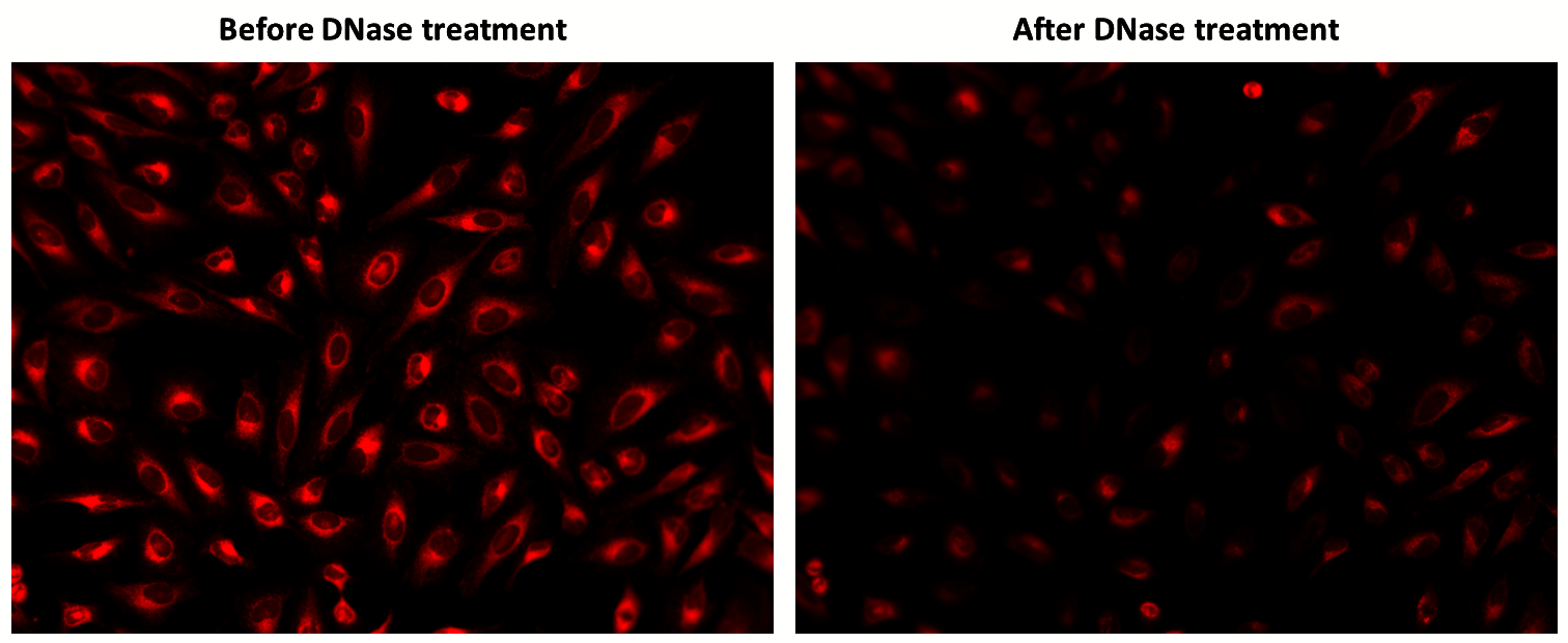
The fluorescence response of MitoDNA™ Red 680 (5 µM) in HeLa cells was assessed before and after DNase treatment. Fluorescence intensities were monitored using fluorescence microscopy.