Mitochondria
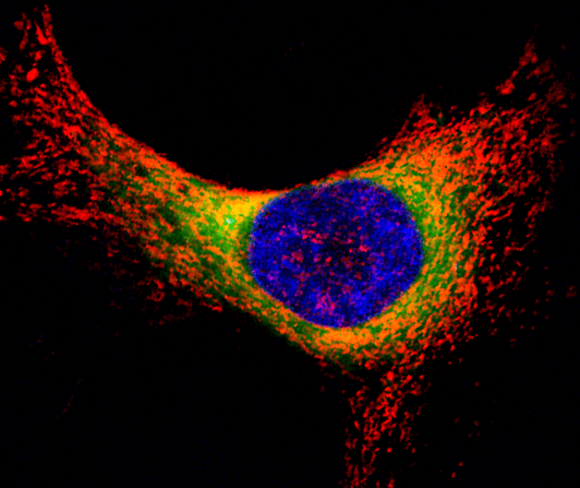
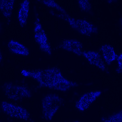
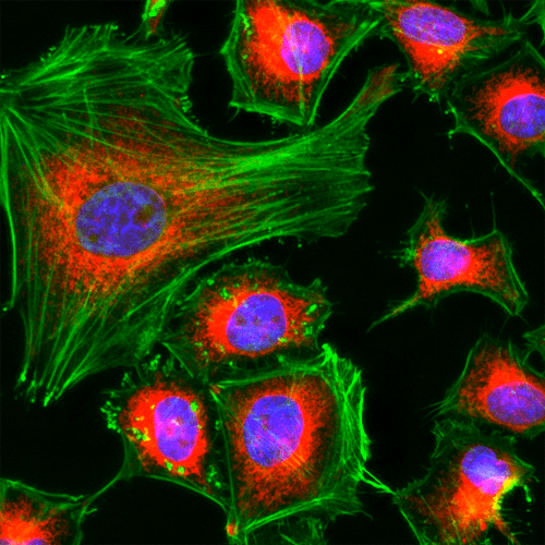
MitoDNA™ Red 610; Mitochondria are a distinctive, membrane-bound organelle present within all eukaryotic cells. As the primary site of ATP production and cellular energy supply, the unique aspects of mitochondria (including separate genetic material termed mDNA, it's own protein synthesizing machinery, and a strictly-regulated membrane potential charge gradient) make it an important subject of study. Additionally, as more aspects of the aging process and other human diseases are being discovered, the role of mitochondria has been shown to be even more prominent than previously suspected.
AAT Bioquest offers a wide range of imaging technologies for investigating mitochondrial morphology and functionality in live and fixed cells. The selective labeling of live cell compartments provides a powerful method for examining cellular events in a spatial and temporal context.
AAT Bioquest offers a wide range of imaging technologies for investigating mitochondrial morphology and functionality in live and fixed cells. The selective labeling of live cell compartments provides a powerful method for examining cellular events in a spatial and temporal context.
Table of Contents
- MitoLite™ Dyes for Live Cell Mitochondrial Imaging
- 1.1 Features of MitoLite™ probes:
- 1.2 CytoFix™ Red Fixable Mitochondrial Stain
- 1.3 Features of CytoFix™ Red mitochondrial stain:
- Mitochondrial Membrane Potential Probes
- 2.1 JC-10™ Dual-Emission ΔΨm Probe
- 2.2 Features of JC-10™:
- 2.3 Mitochondrial-Selective Rhodamine Esters
- MitoDNA™ Dyes for Mitochondrial DNA Labeling
- MitoROS Brite™ Dyes for Mitochondrial ROS Labeling
- Product ordering information
MitoLite™ Dyes for Live Cell Mitochondrial Imaging
MitoLite™ dyes are a series of cell-permeable cationic dyes used to label mitochondria in live cells. At micromolar concentrations (μM), MitoLite™ dyes passively diffuse across live cell membranes and selectively accumulate in active mitochondria via the mitochondrial membrane potential (ΔΨm) gradient. Since staining is lost during cell death due to the collapse (depolarization) of the mitochondrial membrane potential, MitoLite™ dyes can also be used to monitor cell viability. MitoLite™ dyes are available in spectral characteristics ranging from blue to near-infrared to accommodate multiplexing with other fluorescent probes or after fixation for co-localization studies. Retention after fixation varies amongst MitoLite™ dyes, with dyes such as MitoLite™ Blue FX490 being well-retained and MitoLite™ Green FM being lost after fixation. MitoLite™ dyes are key components of Cell Navigator™ Mitochondrion Staining Kits made available separately here. Each kit provides sufficient materials for 500 tests.
Features of MitoLite™ probes:
- Selectively accumulates in active mitochondria via the ΔΨm gradient
- Exceptionally bright fluorescence with high signal-to-noise ratios
- Photostable signal generation for extended imaging windows
- Can be imaged without washing
- Multiple wavelength options for multiplex analysis with other fluorescent probes
- Suitable for both proliferating and non-proliferating cells, as well as, cell in suspension and adherent cells
Mitolite
Table 1. Available MitoLite™ dyes for labeling mitochondria in live cells.
| Product name ▲ ▼ | Sample Type ▲ ▼ | Fixable ▲ ▼ | Ex (nm) ▲ ▼ | Em (nm) ▲ ▼ | Filter Set ▲ ▼ | Unit Size ▲ ▼ | Cat No. ▲ ▼ | Assay Kit No. ▲ ▼ |
| MitoLite™ Blue FX490 | Live Cells | Yes | 344 | 469 | DAPI | 500 Tests | 22674 | 22665 |
| MitoLite™ Green FM | Live Cells | No | 508 | 528 | FITC | 10x50 µg | 22695 | - |
| MitoLite™ Green EX488 | Live Cells | No | 508 | 528 | FITC | 500 Tests | 22675 | 22666 |
| MitoLite™ Orange EX405 | Live Cells | No | 425 | 522 | FITC | 500 Tests | 22679 | 22673 |
| MitoLite™ Orange FX570 | Live Cells | Yes | 553 | 576 | TRITC | 500 Tests | 22676 | 22667 |
| MitoLite™ Red FX600 | Live Cells | Yes | 580 | 598 | TRITC | 500 Tests | 22677 | - |
| MitoLite™ Red CMXRos | Live Cells | Yes | 579 | 599 | TRITC | 10x50 µg | 22698 | 22668 |
| MitoLite™ Deep Red FX660 | Live Cells | Yes | 640 | 659 | Cy5 | 500 Tests | 22678 | 22669 |
| MitoLite™ NIR FX690 | Live Cells | Yes | 658 | 691 | Cy5 | 500 Tests | 22690 | 22670 |
CytoFix™ Red Fixable Mitochondrial Stain
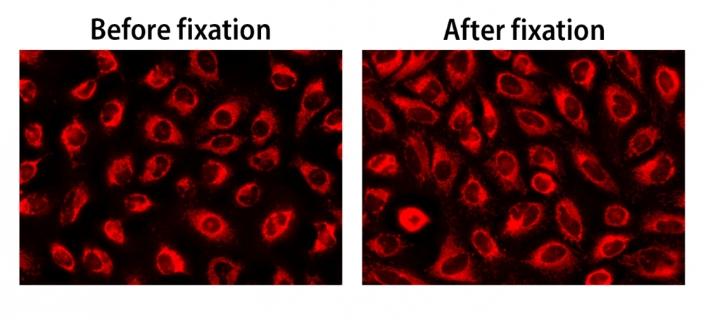
The enhanced cellular retention of CytoFix™ Red mitochondrial stain preserves its mitochondrial localized fluorescence even after the mitochondrial membrane potential is lost during fixation. The fluorescence signal generated by this dye is well-retained in the lyososme making it convenient for long-term mitochondrial tracking. CytoFix™ Red mitochondrial stain can be used with other fluorescent conjugates and other organelle stains for multicolor analysis. It can be used for both suspension and adherent cells and readily adapted for a wide variety of fluorescence platforms.
The fluorescence images of HeLa cells stained with CytoFix™ MitoRed in a 96-well black-wall clear-bottom plate. Image was acquired before (Left) and after (Right) fixation with 4% formaldehyde solution for 20 minutes at RT. The cells were imaged using fluorescence microscope with a Cy3/TRITC filter.
Features of CytoFix™ Red mitochondrial stain:
- High mitochondrial staining efficiency
- Long retention after fixation
- Labeling protocol with minimal hands on time
Table 2. CytoFix™ Red fixable mitochondrial stain
| Product name ▲ ▼ | Sample Type ▲ ▼ | Fixable ▲ ▼ | Ex (nm) ▲ ▼ | Em (nm) ▲ ▼ | Filter Set ▲ ▼ | Unit Size ▲ ▼ | Cat No. ▲ ▼ |
| CytoFix™ Red Mitochondrial Stain | Live Cells | Yes | 540 | 590 | Cy3/TRITC | 500 Tests | 23200 |
Mitochondrial Membrane Potential Probes
The mitochondrial membrane potential (ΔΨm), generated by the electron transport chain, is a key parameter necessary for healthy mitochondrial functioning. Together with the proton gradient, it generates the driving force behind mitochondrial ATP synthesis. It plays a key role in mitochondrial homeostasis through selective elimination of dysfunctional mitochondria, and is an essential component of mitochondrial calcium homeostasis.
A distinctive feature of the early stages of apoptosis is the disruption of normal mitochondrial function. A collapse in mitochondrial membrane and redox potential may induce unwanted loss of cell viability and be a cause of various pathologies. We offer a wide assortment of fluorescent probes for analyzing aspects of normal mitochondrial activity in live cells, including reactive oxygen species (ROS) production, mitochondrial membrane potential and calcium flux.
JC-10™ Dual-Emission ΔΨm Probe
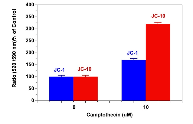
Camptothecin induced mitochondrial membrane potential changes were measured with JC-10™ (Cat No. 22204) and JC-1 (Cat No. 22200) in Jurkat cells. After Jurkat cells were treated with camptothecin (10 μM) for 4 hours, JC-1 and JC-10™ dye loading solutions were added to the wells and incubated for 30 minutes. The fluorescent intensities for both J-aggregates and monomeric forms of JC-1 and JC-10™ were measured at Ex/Em = 490/525 nm and 540/590 nm with NOVOstar microplate reader (BMG Labtech).
Features of JC-10™:
- Easy-to-Use: JC-10™ does not percipitate when diluted into aqueous buffers, eliminating artifacts.
- Robust: JC-10™ has smaller assay deviations due to its enhanced solubility in aqueous media and higher sensitivity.
- Enhanced Signal: JC-10™ has a higher signal-to-background ratio than JC-1.
- Enhanced Sensitivity: JC-10™ has the ability to detect subtle changes in ΔΨm loss better than JC-1 in all tested cell lines.
- Broad Applications: JC-10™ can be used for primary rat hepatocytes.
- Convenient: JC-10™ is compatible with fluorescence microplate readers, cell imagers and flow cytometers.
Table 3. JC-10™ and JC-1 dual-emission mitochondrial membrane potential probes.
| Probe ▲ ▼ | Ex/Em ▲ ▼ | Ex/Em ▲ ▼ | Filter Set ▲ ▼ | Unit Size ▲ ▼ | Cat No. ▲ ▼ |
| JC-10™ *Superior alternative to JC-1* | 508/524 (monomer) | 508/570 (aggregate) | FITC (monomer) TRITC (aggregate) | 5x100 µL | 22204 |
| JC-1 | 515/530 (monomer) | 515/590 (aggregate) | FITC (monomer) TRITC (aggregate) | 5 mg | 22200 |
| JC-1 | 515/530 (monomer) | 515/590 (aggregate) | FITC (monomer) TRITC (aggregate) | 50 mg | 22201 |
Table 4. Cell Meter™ JC-10™ assay kits for measuring mitochondrial membrane potential.
| Probe ▲ ▼ | Instrument ▲ ▼ | Ex (nm) ▲ ▼ | Em (nm) ▲ ▼ | Cutoff/Channel ▲ ▼ | Unit Size ▲ ▼ | Cat No. ▲ ▼ |
| Cell Meter™ JC-10 Mitochondrion Membrane Potential Assay Kit *Optimized for Microplate Assays* | Microplate Reader | 490/540 nm | 525/590 nm | 515/570 nm | 500 tests | 22800 |
| Cell Meter™ JC-10 Mitochondrion Membrane Potential Assay Kit *Optimized for Flow Cytometry Assays* | Flow Cytometer | 488 nm laser | 530/30 575/26 | FITC Channel PE Channel | 100 tests | 22801 |
Mitochondrial-Selective Rhodamine Esters
Cell-permeable cationic rhodamines, such TMRE and TMRM, are readily sequestered by active mitochondria, and commonly used to label mitochondria in living cells. Like JC-10™, TMRE and TMRM uptake in mitochondria is driven by the mitochondrial membrane potential. Both dyes have been successfully for dymanic and in situ quantitative measurements, to screen for inhibitors of the mitochondrial transition pore, to assess the functionality of mitochondria in living cells, and can be used to discrimate between viable and non-viable cell populations. These potentiometric dyes exhibit minimal self-quenching, low cytotoxicity and have reasonable photostability, and their fluorescence intensities can be measured with either a flow cytometer or fluorescence microscope. In comparison to TMRM, TMRE is slightly more hydrophobic.
MitoDNA™ Dyes for Mitochondrial DNA Labeling
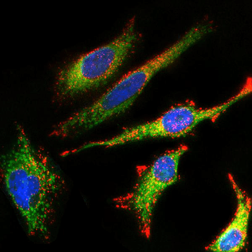
Mitochondrial deoxyribonucleic acid (mtDNA) is small circular DNA found within mitochondria present in the cytoplasm of a cell. This DNA is supplementary to the nucleic acid material found in the nucleus of each cell. The mtDNA codes for 37 genes that promote the proper functioning of some cells. The mitochondria synthesize adenosine triphosphate (ATP) through oxidative phosphorylation and encode information for the synthesis of enzymes, transfer ribonucleic acid (tRNA), and ribosomal RNA (rRNA). Disorders of mtDNA and mutations in its genes can predispose to health problems like age-related hearing loss, diabetes, and brain, heart, and liver failure, among other conditions. Moreover, mtDNA and its associated mitochondrial disorders can predispose people to different types of cancers including lymphomas, leukemias, and breast, intestine, liver and kidney tumors etc. There are very few probes that can be effectively used to detect mtDNA. The common fluorescent DNA probes (such as DAPI, Hoechst or SYBR® Green) lack the specificity to target mitochondria. They predominantly stain nuclei. The MitoDNA™ dye series specifically stain mtDNA in live cells.
Features of MitoDNA™ dyes:
- Cell permeable
- Specific for mtDNA
- Large Stokes Shifts
- Excellent signal-to-noise ratio
- Suitable for multiplex analysis with other fluorescent probes
MitoDNA
MitoROS Brite™ Dyes for Mitochondrial ROS Labeling
Mitochondrial reactive oxygen species (mtROS or mROS), including superoxide anion, hydrogen peroxide and hydroxyl radicals, are primarily generated at the electron transport chain within the inner mitochondrial membrane during oxidative phosphorylation. These reactive species play critical roles in cellular signaling and homeostasis, with their levels tightly regulated to maintain cell function. At basal concentrations, mtROS are involved in the regulation of inflammatory responses and metabolic adaptation. Moderate levels are required for essential cellular processes such as gene expression, cell proliferation, and differentiation. However, excessive mtROS accumulation can activate apoptosis and autophagy pathways, underscoring their involvement in both cell survival and death mechanisms.
Recent advances in cancer research have highlighted the role of mtROS in tumor cell biology. mtROS regulate diverse signaling networks that drive tumorigenesis, including pathways involved in cell proliferation, survival, and metastasis. This has led to the development of novel therapeutic strategies aimed at selectively modulating mtROS levels in cancer cells. In addition to cancer, mtROS are implicated in the pathogenesis of COVID-19. SARS-CoV-2 infection has been shown to increase mtROS production, leading to the upregulation of HIF-1α, a key factor in viral replication. This discovery suggests that targeting mtROS in infected tissues, such as the lungs, could provide a therapeutic avenue for treating COVID-19.
The fluorescence response of MitoROS Brite™ NIR 780 (0.5 µM) to varying concentrations of H2O2 in HeLa cells was assessed. Fluorescence intensities were monitored using a fluorescence microscope equipped with a Cy7 filter.
MitoROS Brite™ dyes are designed for precise and reliable detection of mitochondrial reactive oxygen species (ROS) in live-cell applications. The series includes MitoROS Brite™ 570 and MitoROS Brite™ 670, which target a broad spectrum of ROS, and MitoROS Brite™ NIR 780, which selectively detects hydrogen peroxide. These cell-permeable probes accumulate in mitochondria and are rapidly oxidized by their specific ROS, producing a robust fluorescent signal. These tools are critical for studying oxidative stress and its regulation in diverse pathological conditions, allowing for the clear differentiation between artifacts from isolated mitochondrial systems and direct mtROS measurements in live-cell mitochondria. Additionally, AAT Bioquest provides superoxide-specific dyes, MitoROS™ 520 and MitoROS™ 580 (Cat# 16052), which are key components of the Cell Meter™ Fluorimetric Mitochondrial Superoxide Activity Assay Kits, as well as the hydroxyl radical-selective probe, MitoROS™ OH580, which is the key compoent of the Cell Meter™ Mitochondrial Hydroxl Radical Detection Kit (Cat# 16055). These specialized probes offer enhanced accuracy in assessing mitochondrial ROS dynamics under physiological and pathological conditions.
Features of MitoROS Brite™ dyes:
- Strong retention of oxidized fluorophore within live cells
- Specific for mitochondrial superoxide
- High signal-to-noise ratio
- Optimization for multiplex analysis with other mitochondrial probes
Table 7. Selectivity of MitoROS Brite™ and MitoROS™ dyes for different reactive oxygen species (ROS)
| Reactive Oxygen Species (ROS) ▲ ▼ | MitoROS Brite™ 570 ▲ ▼ | MitoROS Brite™ 670 ▲ ▼ | MitoROS Brite™ NIR 780 ▲ ▼ | MitoROS™ 520 ▲ ▼ | MitoROS™ 580 ▲ ▼ | MitoROS™ OH580 ▲ ▼ |
| Hydrogen peroxide (H2O2) | + | + | +++ | - | - | - |
| Hydroxyl radical (•OH) | ++ | ++ | - | - | - | +++ |
| Tert-butyl-Hydroperoxide (TBHP) | + | + | - | - | - | - |
| Hypochlorous acid (HOCl) | - | + | - | - | - | - |
| Superoxide anion (O2•−) | + | ++ | - | +++ | +++ | - |
| Nitric oxide (NO) | - | - | - | - | - | - |
| Peroxynitrite anion (ONOO−) | - | - | - | - | - | - |
| Cat# | 15998 | 15999 | 16049 | 16060 | 16052 | 16055 |
Product ordering information
Table 9. Ordering Info for Mitochondrial Products
| Cat# ▲ ▼ | Product Name ▲ ▼ | Unit Size ▲ ▼ |
| 67 | Rhodamine 700 *CAS 63561-42-2* | 25 mg |
| 68 | Rhodamine 800 *CAS 137993-41-0* | 25 mg |
| 21510 | OxiVision™ Red Mitochondrial Lipid Peroxidation Sensor | 5x100 µg |
| 21511 | OxiVision™ Ratiometric Mitochondrial Lipid Peroxidation Sensor | 5x100 µg |
| 22205 | Nonyl acridine orange *CAS 75168-11-5* | 25 mg |
| 22210 | Rhodamine 123 *CAS 62669-70-9* | 25 mg |
| 22211 | Rhodamine B, hexyl ester, perchlorate | 10 mg |
| 22220 | TMRE [Tetramethylrhodamine ethyl ester] *CAS#: 115532-52-0* | 25 mg |
| 22221 | TMRM [Tetramethylrhodamine methyl ester] *CAS#: 115532-50-8* | 25 mg |
| 22225 | DASPEI [2-(4-(dimethylamino)styryl)-N-ethylpyridinium iodide] *CAS#: 3785-01-1* | 100 mg |
