MitoLite™ Dyes and Kits
Live Cell Mitochondrial Imaging
MitoLite™ dyes are a series of cell-permeable cationic dyes used to label mitochondria in living cells. At micromolar concentrations (µM), MitoLite™ dyes passively diffuse across live cell membranes and selectively accumulate in active mitochondria via the mitochondrial membrane potential (ΔΨm) gradient. Since staining is lost during cell death due to the collapse (depolarization) of the mitochondrial membrane potential, MitoLite™ dyes can also be used to monitor cell viability. MitoLite™ dyes are available in spectral characteristics ranging from blue to near-infrared to accommodate multiplexing with other fluorescent probes or after fixation for co-localization studies. Retention after fixation varies amongst MitoLite™ dyes, with dyes such as MitoLite™ Blue FX490 being well-retained and MitoLite™ Green FM being lost after fixation. MitoLite™ dyes are key components of Cell Navigator™ Mitochondrion Staining Kits.
Cell Navigator™ Mitochondrion Staining Kits combine the enhanced staining efficiency of MitoLite™ reagents with an optimized staining protocol to rapidly label mitochondria in living cells. Each kit supplies sufficient materials for performing 500 assays. Cell Navigator™ Mitochondrion Staining Kits can be readily adapted for various fluorescence platforms such as microplate assays, immunocytochemistry and flow cytometry. They are effective in a variety of studies, including cell adhesion, chemotaxis, multidrug resistance, cell viability, apoptosis and cytotoxicity.
Features of MitoLite™ probes:
- Selectively accumulates in active mitochondria via the ΔΨm gradient
- Exceptionally bright fluorescence with high signal-to-noise ratios
- Photostable signal generation for extended imaging windows
- Can be imaged without washing
- Multiple wavelength options for multiplex analysis with other fluorescent probes
- Suitable for both proliferating and non-proliferating cells, as well as, cell in suspension and adherent cells
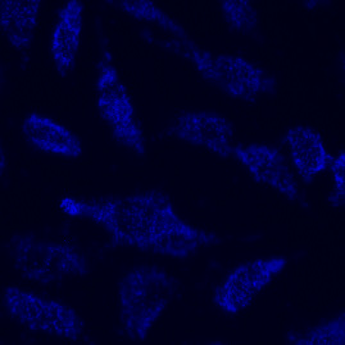
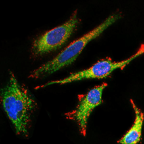
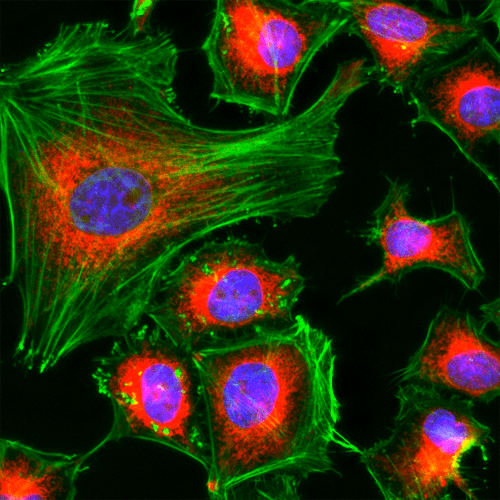
MitoLite™ Images
Table 1. Available MitoLite™ dyes for labeling mitochondria in live cells.
| Product name ▲ ▼ | Sample Type ▲ ▼ | Fixable ▲ ▼ | Ex (nm) ▲ ▼ | Em (nm) ▲ ▼ | Filter Set ▲ ▼ | Unit Size ▲ ▼ | Cat No. ▲ ▼ | Assay Kit No. ▲ ▼ |
| MitoLite™ Blue FX490 | Live Cells | Yes | 344 | 469 | DAPI | 500 Tests | 22674 | 22665 |
| MitoLite™ Green FM | Live Cells | No | 508 | 528 | FITC | 10x50 µg | 22695 | - |
| MitoLite™ Green EX488 | Live Cells | No | 508 | 528 | FITC | 500 Tests | 22675 | 22666 |
| MitoLite™ Orange EX405 | Live Cells | No | 425 | 522 | FITC | 500 Tests | 22679 | 22673 |
| MitoLite™ Orange FX570 | Live Cells | Yes | 553 | 576 | TRITC | 500 Tests | 22676 | 22667 |
| MitoLite™ Red FX600 | Live Cells | Yes | 580 | 598 | TRITC | 500 Tests | 22677 | - |
| MitoLite™ Red CMXRos | Live Cells | Yes | 579 | 599 | TRITC | 10x50 µg | 22698 | 22668 |
| MitoLite™ Deep Red FX660 | Live Cells | Yes | 640 | 659 | Cy5 | 500 Tests | 22678 | 22669 |
| MitoLite™ NIR FX690 | Live Cells | Yes | 658 | 691 | Cy5 | 500 Tests | 22690 | 22670 |
Fluorescence Spectrum Viewer
Need assistance selecting the best fluorophore for your experiment, use our Fluorescence Spectrum Viewer:
- View and compare fluorophores and fluorescent proteins for biological applications
- Check spectral compatibility
- Add multiple excitation and emission filters
- Save spectra configuration as a PNG or hyperlink


