Protonex™ Red 600-Latex Bead Conjugate
Example protocol
AT A GLANCE
Solvent: | Water |
Solids Content: | 1% in PBS |
Number of Microspheres per mL: | ~4e+10 |
Ex/Em: | 575/597 nm |
Mean Diameter: | 0.72 µm |
SAMPLE EXPERIMENTAL PROTOCOL
The following is a recommended protocol for granulocytes. This protocol only provides a guideline and should be modified
according to your specific experimental conditions.
Prepare cells as desired. For example, prepare the granulocytes at 107 cells/mL with Hanks and 20 mM Hepes buffer (HHBS), and add 100 μL to a polypropylene tube.
Note: Each cell line should be evaluated on an individual basis to determine the optimal cell density.
Add 1-10 μL of the Protonex™ Red 600-Latex Bead Conjugate to the tube and incubate with gentle shaking for 30 minutes at 37˚C.
Note: Each cell line should be evaluated on an individual basis to determine the optimal incubation time.
Prepare an identical sample that is incubated at 4˚C and label it as a control.
At the end of the 30-minute incubation, stop phagocytosis by adding 2mL of ice-cold HHBS and mix well.
Wash the cells 2 times with cold HBSS.
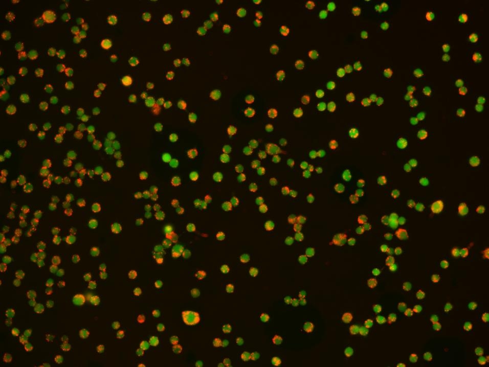
Resuspend the cells in 500 μL of cold HBSS, keep the samples at 4˚C, and analyze immediately using a fluorescence microscope equipped with a Texas Red® filter set.
Note: For fluorescence microplate readers, monitor the fluorescence intensity at Ex/Em = 570/600 nm (Cutoff = 585 nm).
Spectrum

Product family
| Name | Excitation (nm) | Emission (nm) |
| Protonex™ Red 670-Latex Bead Conjugate | 643 | 660 |
Citations
Authors: Lu, Fangfang and Wang, Ronglin and Nie, Tiejian and Gao, Fei and Yang, Shaosong and Huang, Lu and Xia, Li and Shao, Kaifeng and Liu, Jiankang and others,
Journal: (2021)
Authors: Guentsch, Annemarie and Beneke, Angelika and Swain, Lija and Farhat, Katja and Nagarajan, Shunmugam and Wielockx, Ben and Raithatha, Kaamini and Dudek, Jan and Rehling, Peter and Zieseniss, Anke and others,
Journal: Molecular and cellular biology (2017): e00236--16
Authors: Guentsch, Annemarie and Beneke, Angelika and Swain, Lija and Farhat, Katja and Nagarajan, Shunmugam and Wielockx, Ben and Raithatha, Kaamini and Dudek, Jan and Rehling, Peter and Zieseniss, Anke and others, undefined
Journal: Molecular and Cellular Biology (2016): MCB--00236
References
Authors: Sarantis H, Grinstein S.
Journal: Methods Cell Biol (2012): 429
Authors: Schreiner L, Huber-Lang M, Weiss ME, Hohmann H, Schmolz M, Schneider EM.
Journal: J Cell Commun Signal (2011): 135
Authors: Flannagan RS, Grinstein S.
Journal: Methods Mol Biol (2010): 121
Authors: Leclerc L, Boudard D, Pourchez J, Forest V, Sabido O, Bin V, Palle S, Grosseau P, Bernache D, Cottier M.
Journal: Inhal Toxicol (2010): 1091
Authors: Steinberg BE, Grinstein S.
Journal: Methods Mol Biol (2009): 45