Nuclear Green™ Fixable DCS1
5 mM DMSO Solution
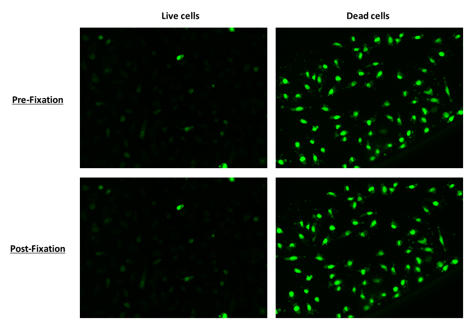
Nuclear Green™ Fixable DCS1 is a fluorogenic, DNA-selective and cell-impermeant dye for analyzing DNA content in dead, fixed or apoptotic cells. It is optimized for surviving cell fixation process better than the common DNA dyes such as PI, DAPI, Hoechst, 7-AAD, DRAQ-5, DRAQ-7 or SYBR Green. It contains a DNA-reactive group, thus covalently bonds with DNA. Nuclear Green™ Fixable DCS1 has its green fluorescence significantly enhanced upon binding to DNA. It can be used in fluorescence imaging, microplate and flow cytometry applications. This DNA-binding dye might be used for multicolor analysis of dead, fixed or apoptotic cells with proper filter sets.


| Catalog | Size | Price | Quantity |
|---|---|---|---|
| 17569 | 0.5 ml | Price |
Physical properties
| Solvent | DMSO |
Spectral properties
| Excitation (nm) | 510 |
| Emission (nm) | 532 |
Storage, safety and handling
| H-phrase | H303, H313, H333 |
| Hazard symbol | XN |
| Intended use | Research Use Only (RUO) |
| R-phrase | R20, R21, R22 |
| Storage | Freeze (< -15 °C); Minimize light exposure |
| UNSPSC | 41116134 |
Instrument settings
| Fluorescence microscope | |
| Excitation | FITC filter set |
| Emission | FITC filter set |
| Recommended plate | Black wall/clear bottom |
Contact us
| Telephone | |
| Fax | |
| sales@aatbio.com | |
| International | See distributors |
| Bulk request | Inquire |
| Custom size | Inquire |
| Technical Support | Contact us |
| Request quotation | Request |
| Purchase order | Send to sales@aatbio.com |
| Shipping | Standard overnight for United States, inquire for international |
Page updated on January 26, 2026

