MycoLight™ Fluorimetric CTC Live Bacteria Quantification Kit
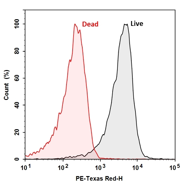
The CTC Flow Cytometric Live Bacteria Assay Kit provides an easy and convenient method for evaluating bacterial health and vitality as a function of the respiratory activity. CTC itself is non-fluorescent, once reduced by the electron transfer system of viable bacterial cell surface, red and insoluble fluorescent CTC formazan is formed and can be detected with a flow cytometer. Dead bacteria that are not respiring or unhealthy bacteria that respire at a lower rate will produce none or less red fluorescent with CTC staining, thus providing a semi-quantitative measure of health status of bacteria population.
Example protocol
AT A GLANCE
Protocol summary
- Prepare 10X dye stock solution
- Prepare bacteria samples
- Incubate bacteria samples with CTC at 37°C for 30 minutes
- Analyze sample by flow cytometry with PE-Texas Red channel
Important
Thaw one of each kit component at room temperature before starting the experiment.
PREPARATION OF STOCK SOLUTION
Unless otherwise noted, all unused stock solutions should be divided into single-use aliquots and stored at -20 °C after preparation. Avoid repeated freeze-thaw cycles.
1. CTC stock solution (10X):
Add 1 mL of distilled water into the vial of CTC (Component A) to make 10X stock solution. Note: Stock solution is stable at -20°C for 2 weeks. Protect from light.
SAMPLE EXPERIMENTAL PROTOCOL
- Prepare bacteria sample with concentration of 106 cells/ml. Grow bacteria into late log phase in appropriate medium. Remove medium by centrifugation at 10,000 x g for 10 minutes and re-suspend the pellet in appropriate amount of PBS. Note: Measure the optical density of the bacterial culture at wavelength = 600 nm (OD600) to determine the cell number. For E. coli culture, OD600 = 1.0 equals 8 x 10⁸ cells/ml.
- Treat cells with test compounds as desired. Remove treatments by centrifugation at 10,000 x g for 10 minutes and re-suspend the pellet in appropriate amount of Assay Buffer (Component B) so the concentration of bacteria in the treated sample is the same as the live. Note: Determine the concentration of the bacterial culture before starting the treatment. Note: Dead bacteria can serve as negative control. It is recommended to kill bacteria with 70% ethanol for 30 min followed by 60 min of boiling.
- Add 10 µL of the 10X CTC stock solution to 90 µL of the bacterial sample.
- Mix well and incubate in the dark for 30 min at 37°C.
- Add 400 µL of Assay Buffer (Component B) to increase volume before analyzing the cells with a flow cytometer.
- Monitor fluorescence of bacteria with a flow cytometer through PE-Taxes Red channel (Ex/Em = 488/615 nm). Note: To exclude debris, it is recommended to set the threshold of the flow cytometer as the following: FSC >10,000, SSC>5,000. Note: The efficiency of CTC is highly strain dependent and the staining conditions would be optimized accordingly.
Citations
View all 25 citations: Citation Explorer
Assessing the effectiveness and mechanisms of Fe/PS, Fe/H2O2, and O3 treatment for water disinfection: Spotlight on VBNC bacteria
Authors: Qi, Zheng and Huang, Zaihui and Liu, Chunguang
Journal: Journal of Environmental Sciences (2024)
Authors: Qi, Zheng and Huang, Zaihui and Liu, Chunguang
Journal: Journal of Environmental Sciences (2024)
Production of Superoxide in Bacteria Is Stress- and Cell State-Dependent: A Gating-Optimized Flow Cytometry Method that Minimizes ROS Measurement Artifacts with Fluorescent Dyes
Authors: McBee, M. E., Chionh, Y. H., Sharaf, M. L., Ho, P., Cai, M. W., Dedon, P. C.
Journal: Front Microbiol (2017): 459
Authors: McBee, M. E., Chionh, Y. H., Sharaf, M. L., Ho, P., Cai, M. W., Dedon, P. C.
Journal: Front Microbiol (2017): 459
Effect of extremely low frequency electromagnetic fields on bacterial membrane
Authors: Oncul, S., Cuce, E. M., Aksu, B., Inhan Garip, A.
Journal: Int J Radiat Biol (2016): 42-9
Authors: Oncul, S., Cuce, E. M., Aksu, B., Inhan Garip, A.
Journal: Int J Radiat Biol (2016): 42-9
Simple and rapid preparation of red fluorescence and red color S. aureus derived nanobioparticles for pathogen detection
Authors: Hu, W., Zhang, Y., Yang, H., Yu, J., Wei, H.
Journal: J Microbiol Methods (2015): 47-53
Authors: Hu, W., Zhang, Y., Yang, H., Yu, J., Wei, H.
Journal: J Microbiol Methods (2015): 47-53
Reduction of biofilm formation on titanium surface with ultraviolet-C pre-irradiation
Authors: Yamada, Y., Yamada, M., Ueda, T., Sakurai, K.
Journal: J Biomater Appl (2014): 161-171
Authors: Yamada, Y., Yamada, M., Ueda, T., Sakurai, K.
Journal: J Biomater Appl (2014): 161-171
Page updated on December 17, 2024