Cell Navigator® Live Cell Endoplasmic Reticulum (ER) Staining Kit *Red Fluorescence*
Example protocol
AT A GLANCE
- Prepare cells in growth medium
- Incubate cells with ER Tracer™ Red working solution at 37 °C for 15 - 30 minutes
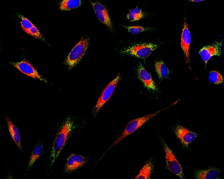
- Analyze under fluorescence microscope at Ex/Em = 590/620 nm (TRITC or Cy3 filter set)
Important Thaw all the kit components at room temperature before starting the experiment.
CELL PREPARATION
For guidelines on cell sample preparation, please visit https://www.aatbio.com/resources/guides/cell-sample-preparation.html
PREPARATION OF STOCK SOLUTIONS
Unless otherwise noted, all unused stock solutions should be divided into single-use aliquots and stored at -20 °C after preparation. Avoid repeated freeze-thaw cycles
Add 20 µL of DMSO (Component C) into the vial of ER Tracer™ Red (Component A) and mix well to make 500X ER Tracer™ Red stock solution.
Note 20 µL of 500X ER Tracer™ Red stock solution is enough for one 96-well plate. Unused 500X ER Tracer™ Red stock solution can be stored at ≤ -20 ºC for two weeks if the tubes are sealed tightly. Protect from light.
PREPARATION OF WORKING SOLUTION
Add 20 µL of 500X ER Tracer™ Red stock solution into 10 mL of Live Cell Staining Buffer (Component B), and mix well to make ER Tracer™ Red working solution.
Note This ER Tracer™ Red working solution is stable for at least 2 hours at room temperature. Protect from light.
SAMPLE EXPERIMENTAL PROTOCOL
- Plate and treat cells as desired.
- Remove cell culture medium. Optional: Cells can be washed twice with buffer of your choice.
Add 100 µL/well (96-well plate) or 50 µL/well (384-well plate) of ER Tracer™ Red working solution in the cell plate. Incubate cells with working solution at 37 °C for 15 - 30 minutes, protected from light.
Note The optimal concentration of the ER probe varies depending on the specific application. Concentration higher than the working solution can be toxic to cells. The staining conditions may be modified according to the particular cell type and the permeability of the cells or tissues to the probe.
Remove ER Tracer™ Red working solution in each well. Wash cells with physically relevant buffer three times.
Fix cells after staining (Optional). Fix the cells with 4% formaldehyde for 5 - 10 minutes. Wash cells with physically relevant buffer three times.
Observe the fluorescence signal in cells using fluorescence microscope with TRITC or Cy3 filter set (Ex/Em = 590/620 nm).
Spectrum

Product family
| Name | Excitation (nm) | Emission (nm) |
| Cell Navigator® Live Cell Endoplasmic Reticulum (ER) Staining Kit *Blue Fluorescence* | 344 | 457 |
| Cell Navigator® Live Cell Endoplasmic Reticulum (ER) Staining Kit *Green Fluorescence* | 503 | 511 |
Citations
Authors: Huang, Haoyang and Liu, Shaowei and Xu, Ligeng and Liang, Hengrui and Wu, Zihao and Chen, Tianfeng and Wang, Jinlin and Liu, Jun
Journal: Journal of Nanobiotechnology (2025): 294
Authors: Hirata, Yoko and Takemori, Hiroshi and Furuta, Kyoji and Kamatari, Yuji O and Sawada, Makoto
Journal: Current Research in Pharmacology and Drug Discovery (2024): 100196
Authors: Zhang, Jun and Peng, Yu and Song, Haosen and Liu, Siqi and Li, Chuanwei and Zhang, Yi and Shi, Xiaowei and Guo, Huifang and Xu, Yingping
Journal: International Immunopharmacology (2024): 112012