TUNEL Assay
Green Fluorescence
Cell Meter™ Fixed Cell and Tissue TUNEL Apoptosis Assay Kit provides a robust tool for conveniently detecting DNA fragmentation caused by apoptosis. The assay is a non-radioactive, simple, accurate and rapid method for monitoring apoptosis in. fixed cells and tissues via imaging DNA fragmentation. The TUNEL assay uses terminal deoxynucleotidyl transferase (TdT) to catalyze the incorporation of fluorescein-12-dUTP at the 3’-hydroxyl ends of the fragmented DNA. The fluorescein-labeled DNA is analyzed by fluorescence microscopy or flow cytometry (excitation at 488 nm with 530/30 nm emission filter). The kit can be used to detect apoptosis in fixed cells and formalin-fixed, paraffin-embedded tissue sections.
Example protocol
AT A GLANCE
Protocol summary
- Treat samples as desired
- Fix cells with 4% formaldehyde solution for 30 minutes on ice
- Permeabilize cells with 70% ice-cold ethanol for 60 minutes on ice
- Add TdT staining solution to samples and incubate for 60 minutes at 37 °C
- Monitor the fluorescence intensity using fluorescence microscopy with FITC filter set
Important
Bring all the kit components at room temperature before starting the experiment.PREPARATION OF WORKING SOLUTION
TdT staining solution
For one test, Mix the following to make a total volume of 51 µL;45 µL TdT Reaction Buffer (Component D)
5 µL CoCl2 (Component C)
0.5 µL Fluorescein 12-dUTP (Component B)
0.5 µL TdT enzyme (Component A).
Note TdT staining solution should be used promptly.
SAMPLE EXPERIMENTAL PROTOCOL
Protocol for cells staining
The following protocol can be used as a guideline and should be optimized according to the needs.- Treat your samples as desired.
- Wash the samples with buffer of your choice such as PBS containing Ca+2 and Mg+2.
- Fix the samples by adding 100 µL of 4% paraformaldehyde in PBS and incubate the samples for 30 minutes on ice.
- Remove fixation solution and wash samples with PBS.
- Add 100 µL of 70% of ice cold ethanol to samples and incubate the samples for 60 minutes on ice.
Note Samples can be stored at -20 °C at this step for several days before use. - Remove alcohol and wash cells with PBS.
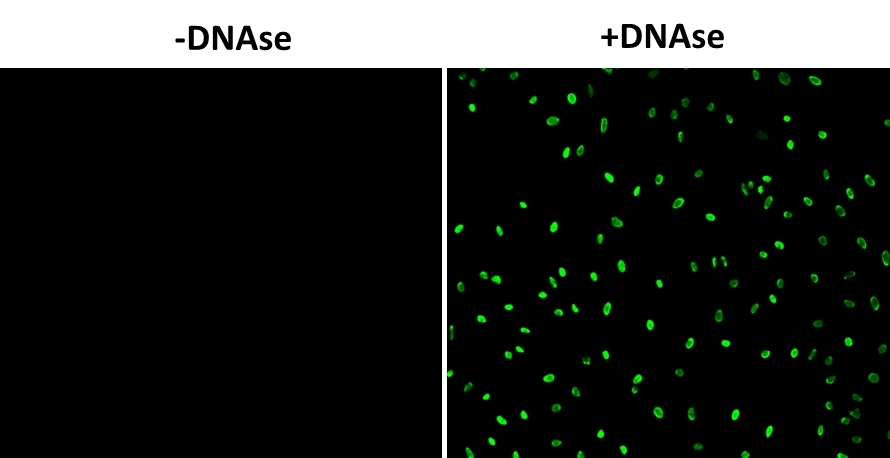
Note For a positive control, incubate fixed samples with 2-5 µg/mL of DNAse in PBS containing Ca+2 and Mg+2 for 60 minutes at 37 °C. Remove the DNAse and wash cells thoroughly and continue with the rest of the protocol - Add 50 µL of TdT staining solution to the samples and incubate for 60 to 120 minutes at 37 °C.
- Remove TdT working solution and wash samples with PBS.
- Resuspend the samples in PBS and monitor the fluorescence intensity with flow cytometer using 530 /30 nm filter (FITC channel) or fluorescence microscope with FITC filter set.
Protocol for tissue staining
The following protocol can be used as a guideline and should be optimized according to the needs.Deparaffinization and rehydration protocol
- Deparaffinize tissue sections (attached to the microscopic slides) by immersing slides in fresh xylene in a Coplin jar for 5 minutes at room temperature. Repeat one more time. (Total 2 washes)
- Wash the samples by immersing the slides in 100% ethanol for 5 minutes at room temperature in a Coplin jar.
- Rehydrate the samples by immersing the slides through various concentrations of alcohol subsequently (100, 95, 85, 70, 50%) for 5 minutes each at room temperature
- Wash the samples by immersing the slides in 0.85% NaCl for 5 minutes at room temperature.
- Wash the samples by immersing the slides in PBS for 5 minutes at room temperature. Repeat one more wash. (Total 2 washes)
- Fix the tissue sections by immersing slides in 4% paraformaldehyde solution in PBS for 15-20 minutes at room temperature.
- Wash the samples by immersing the slides in PBS for 5 minutes at room temperature. Repeat one more wash. (Total 2 washes)
- Remove the liquid and place the slides on a flat surface. Treat tissue sections with 100 µL of 20 µg/mL Proteinase K solution. Add enough to cover the entire tissue surface. Incubate slides for 10 minutes at room temperature.
- Wash the samples by immersing the slides in PBS for 5 minutes at room temperature.
- Fix the tissue sections by immersing slides in 4% paraformaldehyde solution in PBS for 15-20 minutes at room temperature.
- Wash the samples by immersing the slides in PBS for 5 minutes at room temperature. Repeat one more wash. (Total 2 washes)
- Optional: For a positive control, incubate fixed samples with 2-5 µg/mL of DNAse in PBS containing Ca+2 and Mg+2 for 60 minutes at 37 °C. Remove the DNAse and wash cells thoroughly with PBS and continue with the rest of the protocol.
- Add 50 µL of TdT staining solution to the samples and incubate for 60 to 120 minutes at 37 °C.
- Remove TdT working solution and wash samples with PBS.
- Add mounting medium with DAPI (AAT Bioquest Cat# 20005) and monitor the fluorescence intensity fluorescence microscope with FITC filter set.
Spectrum

Alternative formats
| Name | Sample Type | Fluorescence |
| Cell Meter™ Live Cell TUNEL Apoptosis Assay Kit *Green Fluorescence* | Live Cell | Green |
| Cell Meter™ Live Cell TUNEL Apoptosis Assay Kit *Red Fluorescence* | Live Cell | Red |
| Cell Meter™ Fixed Cell and Tissue TUNEL Apoptosis Assay Kit *Green Fluorescence* | Fixed Cell and Tissue | Green |
| Cell Meter™ Fixed Cell and Tissue TUNEL Apoptosis Assay Kit *Red Fluorescence* | Fixed Cell and Tissue | Red |
| Cell Meter™ Fixed Cell and Tissue TUNEL Apoptosis Assay Kit *Deep Red Fluorescence* | Fixed Cell and Tissue | Deep Red |
| Cell Meter™ Fixed Cell and Tissue TUNEL Apoptosis Assay Kit *Blue Fluorescence* | Fixed Cell and Tissue | Blue |
Product family
| Name | Excitation (nm) | Emission (nm) | Extinction coefficient (cm -1 M -1) | Correction Factor (260 nm) | Correction Factor (280 nm) |
| Cell Meter™ Fixed Cell and Tissue TUNEL Apoptosis Assay Kit *Blue Fluorescence* | 411 | 472 | - | 0.14 | 0.12 |
| Cell Meter™ Fixed Cell and Tissue TUNEL Apoptosis Assay Kit *Red Fluorescence* | 544 | 570 | 100000 | 0.27 | 0.34 |
| Cell Meter™ Fixed Cell and Tissue TUNEL Apoptosis Assay Kit *Deep Red Fluorescence* | 649 | 663 | 250000 | - | 0.027 |
Citations
View all 9 citations: Citation Explorer
Immuno-protective vesicle-crosslinked hydrogel for allogenic transplantation
Authors: Wang, Yuqian and Huang, Renqi and Lu, Yougong and Liu, Mingqi and Mo, Ran
Journal: Nature Communications (2024): 1--13
Authors: Wang, Yuqian and Huang, Renqi and Lu, Yougong and Liu, Mingqi and Mo, Ran
Journal: Nature Communications (2024): 1--13
In Vitro Effects of Boric Acid on Cell Cycle, Apoptosis, and miRNAs in Medullary Thyroid Cancer Cells
Authors: Y{\i}ld{\i}r{\i}m, Onurcan and Se{\c{c}}me, M{\"u}cahit and Dodurga, Yavuz and Mete, G{\"u}l{\c{c}}in Abban and Fenkci, Semin Melahat
Journal: Biological Trace Element Research (2024): 1--11
Authors: Y{\i}ld{\i}r{\i}m, Onurcan and Se{\c{c}}me, M{\"u}cahit and Dodurga, Yavuz and Mete, G{\"u}l{\c{c}}in Abban and Fenkci, Semin Melahat
Journal: Biological Trace Element Research (2024): 1--11
Antifungal Activity of Cedrol from Cunninghamia lanceolate var. konishii against Phellinus noxius and Its Mechanism
Authors: Hsiao, Wen-Wei and Lau, Ka-Man and Chien, Shih-Chang and Chu, Fang-Hua and Chung, Wen-Hsin and Wang, Sheng-Yang
Journal: Plants (2024): 321
Authors: Hsiao, Wen-Wei and Lau, Ka-Man and Chien, Shih-Chang and Chu, Fang-Hua and Chung, Wen-Hsin and Wang, Sheng-Yang
Journal: Plants (2024): 321
Antiapoptotic and antioxidant effects of melatonin on cat vitrified oocytes
Authors: Colombo, M and Mascaro, A and Pecile, A and Fusi, J and Luvoni, GC and others,
Journal: REPRODUCTION IN DOMESTIC ANIMALS (2023): 192--192
Authors: Colombo, M and Mascaro, A and Pecile, A and Fusi, J and Luvoni, GC and others,
Journal: REPRODUCTION IN DOMESTIC ANIMALS (2023): 192--192
Effects of boric acid on invasion, migration, proliferation, apoptosis and miRNAs in medullary thyroid cancer cells
Authors: Y{\i}ld{\i}r{\i}m, Onurcan and Se{\c{c}}me, M{\"u}cahit and Dodurga, Yavuz and Mete, G{\"u}l{\c{c}}in Abban and Fenkci, Semin Melahat
Journal: (2023)
Authors: Y{\i}ld{\i}r{\i}m, Onurcan and Se{\c{c}}me, M{\"u}cahit and Dodurga, Yavuz and Mete, G{\"u}l{\c{c}}in Abban and Fenkci, Semin Melahat
Journal: (2023)
References
View all 50 references: Citation Explorer
Improvement of in situ Follicular Activation and Early Development in Cryopreserved Human Ovarian Cortical Tissue by Co-Culturing with Mesenchymal Stem Cells.
Authors: Hosseini, Marzieh and Salehpour, Saghar and Ghaffari Novin, Marefat and Shams Mofarahe, Zahra and Abdollahifar, Mohammad-Amin and Piryaei, Abbas
Journal: Cells, tissues, organs (2020): 1-11
Authors: Hosseini, Marzieh and Salehpour, Saghar and Ghaffari Novin, Marefat and Shams Mofarahe, Zahra and Abdollahifar, Mohammad-Amin and Piryaei, Abbas
Journal: Cells, tissues, organs (2020): 1-11
Exosomes derived from adipose tissue, bone marrow, and umbilical cord blood for cardioprotection after myocardial infarction.
Authors: Xu, Huiyu and Wang, Zhongchao and Liu, Longmei and Zhang, Baoxia and Li, Bao
Journal: Journal of cellular biochemistry (2020): 2089-2102
Authors: Xu, Huiyu and Wang, Zhongchao and Liu, Longmei and Zhang, Baoxia and Li, Bao
Journal: Journal of cellular biochemistry (2020): 2089-2102
Vitrification freezing of large ovarian tissue in the human body.
Authors: Zhao, Qian and Zhang, Ying and Su, Ke and Wang, Xiao-Wan and Hai, Pan-Pan and Han, Bing and Bian, Ai-Ping and Guo, Rui-Xia
Journal: Journal of ovarian research (2019): 77
Authors: Zhao, Qian and Zhang, Ying and Su, Ke and Wang, Xiao-Wan and Hai, Pan-Pan and Han, Bing and Bian, Ai-Ping and Guo, Rui-Xia
Journal: Journal of ovarian research (2019): 77
Systemic LPS induces toll-like receptor 3 (TLR3) expression and apoptosis in testicular mouse tissue.
Authors: Nejsum, Lene N and Piec, Adrian and Fijak, Monika and Ernstsen, Christina V and Fischer, Dania and Maier, Thorsten J and Kinscherf, Ralf and Hofmann, Rainer and Urbschat, Anja
Journal: Cell and tissue research (2019): 143-154
Authors: Nejsum, Lene N and Piec, Adrian and Fijak, Monika and Ernstsen, Christina V and Fischer, Dania and Maier, Thorsten J and Kinscherf, Ralf and Hofmann, Rainer and Urbschat, Anja
Journal: Cell and tissue research (2019): 143-154
Classification, Scoring, and Quantification of Cell Death in Tissue Sections.
Authors: Janke, Laura J and Ward, Jerrold M and Vogel, Peter
Journal: Veterinary pathology (2019): 33-38
Authors: Janke, Laura J and Ward, Jerrold M and Vogel, Peter
Journal: Veterinary pathology (2019): 33-38
Page updated on October 8, 2024