logo
AAT Bioquest

Calcein Deep Red™ acetate

This product is now replaced by #22011. Calcein AM is one the most popular fluorescent probes used for labeling and monitoring cellular functions of live cells. However, the single color of Calcein AM makes it impossible to use this valuable reagent in the multicolor applications. For example, it is impossible to use Calcein AM in combination of GFP-tranfacted cells due to the same color to GFP. To address this color limitation of Calcein AM, we have developed Calcein Orange™, Calcein Red™ and Calcein Deep Red™. These new Calcein AM analogs enable the multicolor labeling and functional analysis of live cells in combination with Calcein AM. Non-fluorescent Calcein Deep Red™ acetate can easily get into live cells and hydrolyzes to generate strongly fluorescent Calcein Deep Red™ (Cat#: 21902) dye. Calcein Deep Red™ dye can be monitored with the common Cy5 filter set.
Example protocol

PREPARATION OF STOCK SOLUTIONS

Unless otherwise noted, all unused stock solutions should be divided into single-use aliquots and stored at -20 °C after preparation. Avoid repeated freeze-thaw cycles.

Calcein Deep Red™ Acetate Stock Solution
Prepare a 2 to 5 mM stock solution of Calcein Deep Red™ acetate in high-quality, anhydrous DMSO.
Note     The nonionic detergent Pluronic® F-127 can be used to increase the aqueous solubility of AM esters. In the staining buffer, the final Pluronic® F-127 concentration should be approximately 0.02%. A variety of Pluronic® F-127 products can be purchased from AAT Bioquest. Avoid long-term storage of AM esters in the presence of Pluronic® F-127.

PREPARATION OF WORKING SOLUTION

Calcein Deep Red™ Acetate Working Solution
Prepare a Calcein Deep Red™ acetate working solution of 1 to 10 µM in the buffer of your choice (e.g., Hanks and Hepes buffer). For most cell lines, Calcein Deep Red™ acetate at the final concentration of 4 to 5 µM is recommended. The exact concentration of indicators required for cell loading must be determined empirically.
Note     If your cells contain organic anion-transporters, probenecid (1–2.5 mM) or sulfinpyrazone (0.1–0.25 mM) may be added to the working solution to reduce leakage of the de-esterified indicators.

SAMPLE EXPERIMENTAL PROTOCOL

  1. Prepare cells for imaging.
  2. Remove the cell culture medium and wash cells once with serum-free buffer to remove any remaining media.
    Note     Serum in cell culture media may contain esterase activity, which can increase background interference.
  3. Add Calcein Deep Red™ acetate working solution to the culture.
  4. Incubate cells at 37 °C for 30 to 60 minutes.
  5. Replace the dye working solution with HHBS or buffer of your choice (containing an anion transporter inhibitor, such as 1 mM probenecid, if applicable) to remove any excess probes.
  6. Measure the fluorescence intensity using either a fluorescence microscope equipped with a Cy5 filter set, a flow cytometer equipped with a 660/20 nm filter (APC channel), or a fluorescence plate reader at Ex/Em = 620/660 nm cutoff 630 nm. 

Calculators

Common stock solution preparation

Table 1. Volume of DMSO needed to reconstitute specific mass of Calcein Deep Red™ acetate to given concentration. Note that volume is only for preparing stock solution. Refer to sample experimental protocol for appropriate experimental/physiological buffers.

0.1 mg0.5 mg1 mg5 mg10 mg
1 mM188.551 µL942.756 µL1.886 mL9.428 mL18.855 mL
5 mM37.71 µL188.551 µL377.102 µL1.886 mL3.771 mL
10 mM18.855 µL94.276 µL188.551 µL942.756 µL1.886 mL

Molarity calculator

Enter any two values (mass, volume, concentration) to calculate the third.

Mass (Calculate)Molecular weightVolume (Calculate)Concentration (Calculate)Moles
/=x=

Spectrum

Citations

View all 20 citations: Citation Explorer
Radioiodination of extravesicular surface constituents to study the biocorona, cell trafficking and storage stability of extracellular vesicles
Authors: Yerneni, Saigopalakrishna S and Solomon, Talia and Smith, Jason and Campbell, Phil G
Journal: Biochimica et Biophysica Acta (BBA)-General Subjects (2022): 130069
Bioprinting Exosome-Like Extracellular Vesicle Microenvironments
Authors: Yerneni, Saigopalakrishna S and Whiteside, Theresa L and Weiss, Lee E and Campbell, Phil G
Journal: Bioprinting (2019): e00041
Isoastragaloside I suppresses LPS-induced tight junction disruption and monocyte adhesion on bEnd. 3 cells via an activating Nrf2 antioxidant defense system
Authors: Li, Hong-Li and Jin, Jin-Mei and Yang, Chun and Wang, Ping and Huang, Fei and Wu, Hui and Zhang, Bei-Bei and Shi, Hai-Lian and Wu, Xiao-Jun
Journal: RSC advances (2018): 464--471
Functional imaging of neuronal activity of auditory cortex by using Cal-520 in anesthetized and awake mice
Authors: Li, Jingcheng and Zhang, Jianxiong and Wang, Meng and Pan, Junxia and Chen, Xiaowei and Liao, Xiang
Journal: Biomedical Optics Express (2017): 2599--2610
NINJ2--A novel regulator of endothelial inflammation and activation
Authors: Wang, Jingjing and Fa, Jingjing and Wang, Pengyun and Jia, Xinzhen and Peng, Huixin and Chen, Jing and Wang, Yifan and Wang, Chenhui and Chen, Qiuyun and Tu, Xin and others, undefined
Journal: Cellular Signalling (2017)

References

View all 84 references: Citation Explorer
Functional evidence that the self-renewal gene NANOG regulates esophageal squamous cancer development
Authors: Li, Deng and Xiang, Xiaocong and Yang, Fei and Xiao, Dongqin and Liu, Kang and Chen, Zhu and Zhang, Ruolan and Feng, Gang
Journal: Biochemical and Biophysical Research Communications (2017)
Localized functional chemical stimulation of TE 671 cells cultured on nanoporous membrane by calcein and acetylcholine
Authors: Zibek S, Stett A, Koltay P, Hu M, Zengerle R, Nisch W, Stelzle M.
Journal: Biophys J. (2006)
A vaccination and challenge model using calcein marked fish
Authors: Klesius PH, Evans JJ, Shoemaker CA, Pasnik DJ.
Journal: Fish Shellfish Immunol (2006): 20
Novel fluorescence assay using calcein-AM for the determination of human erythrocyte viability and aging
Authors: Bratosin D, Mitrofan L, Palii C, Estaquier J, Montreuil J.
Journal: Cytometry A (2005): 78
Cytotoxic effects of 100 reference compounds on Hep G2 and HeLa cells and of 60 compounds on ECC-1 and CHO cells. I mechanistic assays on ROS, glutathione depletion and calcein uptake
Authors: Schoonen WG, Westerink WM, de Roos JA, Debiton E.
Journal: Toxicol In Vitro (2005): 505
Page updated on February 1, 2023

Ordering information

Price
Unit size
Catalog Number22010
Quantity
Add to cart

Additional ordering information

Telephone1-800-990-8053
Fax1-800-609-2943
Emailsales@aatbio.com
InternationalSee distributors
Bulk requestInquire
Custom sizeInquire
Technical SupportContact us
Purchase orderSend to sales@aatbio.com
ShippingStandard overnight for United States, inquire for international
Request quotation

Physical properties

Molecular weight

530.36

Solvent

DMSO

Spectral properties

Excitation (nm)

643

Emission (nm)

663

Storage, safety and handling

H-phraseH303, H313, H333
Hazard symbolXN
Intended useResearch Use Only (RUO)
R-phraseR20, R21, R22

Storage

Freeze (< -15 °C); Minimize light exposure
UNSPSC12352200

Platform

Flow cytometer

Excitation640 nm laser
Emission660, 20 nm filter
Instrument specification(s)APC channel

Fluorescence microscope

ExcitationCy5 filter set
EmissionCy5 filter set
Recommended plateBlack wall, clear bottom

Fluorescence microplate reader

Excitation620
Emission660
Cutoff630
Recommended plateSolid black
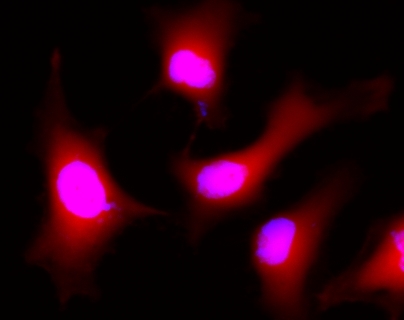
Images of Live HeLa cells stained with Calcein Deep Red&trade; acetate (Cat.22010 ). Cell nuclei were stained with Hoechst 33342 (Blue, Cat#17535).
Fluorescence images of HeLa cells stained with Calcein Deep Red&trade; acetate in a Costar black wall/clear bottom 96-well plate.