Cell Viability Assays
Cell viability assays use a variety of markers to accurately determine live, healthy cells within complex populations. They are used extensively in cytotoxicity tests and drug discovery to measure the health of cells in response to extracellular stimuli, evaluate the efficacy of novel therapeutics, and the sensitivity of cell lines to specific agents. More importantly, viability assays optimize growth and experimental conditions in cell culture to ensure high-quality data that is robust and reproducible.
AAT Bioquest offers a broad range of colorimetric, fluorescence- and bioluminescence-based viability assays and reagents for detecting parameters unique to live, metabolically active cells. These include assays for measuring membrane integrity, enzymatic and metabolic activity, ATP concentration, and mitochondrial membrane potential. In addition to cell viability assays for single-parameter readouts, we offer Live or Dead™ Cell Viability Assays to simultaneously measure live and dead cells based on membrane integrity and metabolic activity.
AAT Bioquest offers a broad range of colorimetric, fluorescence- and bioluminescence-based viability assays and reagents for detecting parameters unique to live, metabolically active cells. These include assays for measuring membrane integrity, enzymatic and metabolic activity, ATP concentration, and mitochondrial membrane potential. In addition to cell viability assays for single-parameter readouts, we offer Live or Dead™ Cell Viability Assays to simultaneously measure live and dead cells based on membrane integrity and metabolic activity.
Table of Contents
- Overview of Cell Viability Assays
- Measuring Plasma Membrane Integrity
- 2.1 Trypan Blue Dye Exclusion Assays
- 2.2 Nucleic Acid Viability Dyes
- 2.3 Propidium Iodide, 7-Aminoactinomycin D, and Cyanine Dyes for Measuring Dead Cells
- 2.4 PI and 7-AAD
- 2.5 Dimeric Cyanine Dyes
- 2.6 Nuclear DCS1™ Dead Cell Stains
- 2.7 Amine-Reactive Cell Meter™ Fixable Viability Dyes
- Measuring Enzymatic Activity in Live Cells
- 3.1 Calcein AM Dyes for Labeling Live Cells
- 3.2 CytoCalcein™ Violet Viability Dyes Optimized for Flow Cytometry
- Measuring Metabolic Activity in Live Cells
Overview of Cell Viability Assays
Table 1. Overview of Cell Viability Assays
| Parameter ▲ ▼ | Assay/Reagents ▲ ▼ | Mode ▲ ▼ | Instrument ▲ ▼ | Principle ▲ ▼ |
| Membrane Integrity | Trypan Blue vital stains | Colorimetric | Hemocytometer | Trypan Blue, Trypan UltraBlue™, Trypan Red Plus™, and Trypan Purple™ are membrane-impermeant vital stains. Live cells with uncompromised membrane integrity exclude these dyes and remain unstained, whereas dead cells do not. The percentage of viable cells equals the number of unstained cells divided by the total number of cells (stained and unstained) multiplied by 100. |
| Membrane Integrity | Membrane-Impermeant DNA dyes | Fluorescence | FC, FM, or MR | Cell-permeable DNA dyes, such as propidium iodide, 7-AAD, or Nuclear DCS1™ dyes, enter dead cells with compromised membranes, whereby they bind to DNA and fluoresce brightly. Viable cells exclude these dyes and remain unstained. |
| Membrane Integrity | Cell Meter™ Fixable Viability dyes | Fluorescence | FC or FM | Cell-impermeable, amine-reactive dyes: in live cells, Cell Meter™ Fixable Viability dyes bind to primary amines on cell surface proteins amine and fluoresce dimly. In dead cells with damaged membranes, these dyes enter the cytoplasm and bind to many intracellular amine groups, resulting in significant fluorescence. The fluorescence intensity difference allows you to discriminate between live and dead cells. |
| Enzymatic activity | Calcein AM dyes | Fluorescence | FC, FM, or MR | Cell-permeable fluorogenic esterase substrates for identifying live cells. These substrates diffuse freely and noninvasively across the plasma membrane of live cells. Once inside, cytosolic esterases in active cells rapidly hydrolyze the non-fluorescent calcein AM substrates into brightly fluorescent calcein products retained by live cells with intact plasma membranes. The fluorescence intensity is proportional to the number of viable cells. |
| Metabolic activity | Tetrazolium dyes (WST and MTT) | Colorimetric | MR | Chromogenic substrates measure cellular reduction potential in metabolically active cells to evaluate viability. Active dehydrogenases and reductases in live, healthy cells reduce tetrazolium dyes from a colorless complex to a brightly purple-colored formazan product. Absorbance is measured at OD = 570 nm to determine viability. |
| Metabolic activity | Resazurin | Colorimetric or fluorescence | MR | Resazurin is an oxidation-reduction indicator. In metabolically active cells, resazurin is reduced by aerobic respiration to resorufin, a pink and highly fluorescent product. The signal produced by resorufin is proportional to the number of viable cells and can be measured using fluorescent or colorimetric instruments. |
| ATP concentration | PhosphoWorks™ ATP assays | Colorimetric, fluorescence, or luminescence | MR | PhosphoWorks™ Luminometric ATP assays take advantage of the luciferin-luciferase reaction, which uses ATP to produce photons of light. The intensity of the luminescent signal is proportional to the concentration of ATP and the number of viable cells. The PhosphoWorks™ Colorimetric ATP assay and PhosphoWorks™ Fluorimetric ATP assay use serial ATP-induced enzyme coupled reactions to produce hydrogen peroxide, which is spectrophotometrically quantified using either a chromogenic or fluorgenic substrate, respectively. In both scenarios, the intensity of the signal is proportional to the concentration of ATP and the number of viable cells. |
| ATP concentration | ReadiUse™ ATP assays | Luminescence | MR | ReadiUse™ Luminometric ATP assays take advantage of the luciferin-luciferase reaction, which uses ATP to produce photons of light. The intensity of the luminescent signal is proportional to the ATP concentration and the number of viable cells. |
- FC = flow cytometer; FM = fluorescence microscope; MR = microplate reader
Measuring Plasma Membrane Integrity
Plasma membrane integrity is the most widely measured parameter for determining cell viability. These methods rely on the principle that viable cells have intact plasma membranes and are impermeable to polar dyes, whereas dead cells are not. The compromised membrane integrity of dead cells allows these dyes to penetrate the membrane and stain intracellular components, such as nucleic acids and proteins. Stained dead cells can then be identified and removed from the final analysis by gating on the unstained, live cell population. Membrane integrity dyes are simple-to-use, reliable, and can be combined with intracellular esterase substrates, membrane potential-sensitive dyes, and organelle probes for multiplex analysis. Common membrane integrity dyes include trypan blue vital stains, nucleic acid binding dyes, and amine-reactive viability dyes.
Trypan Blue Dye Exclusion Assays
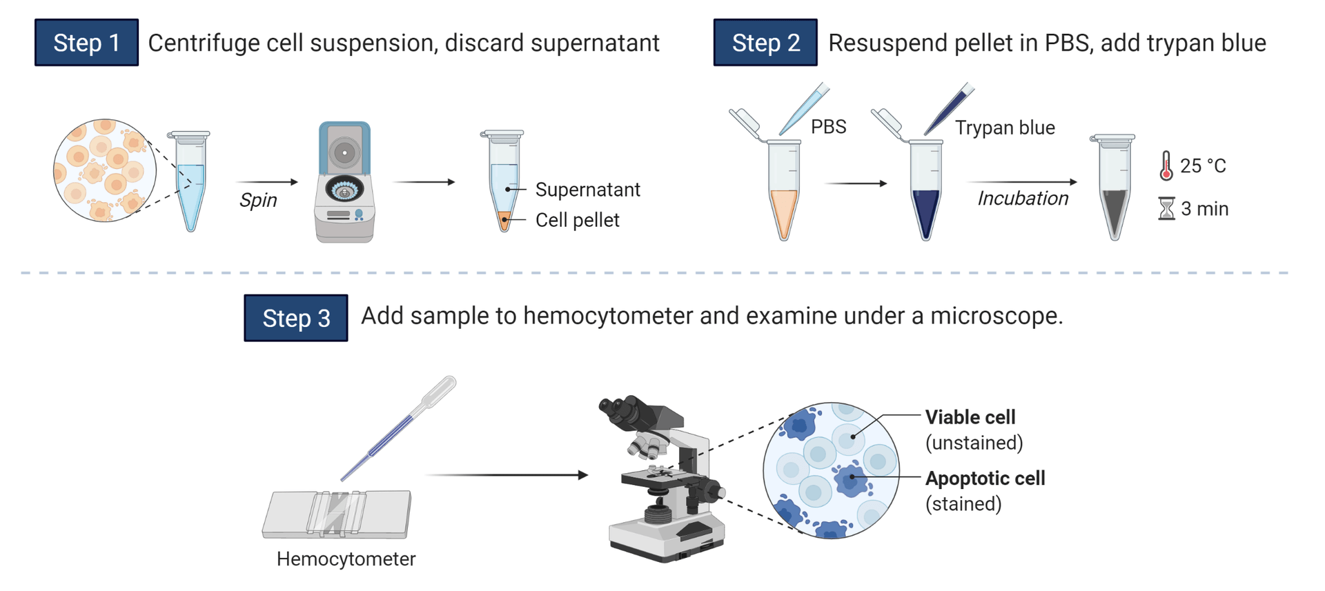
The trypan blue dye exclusion assay is one of the earliest and most common methods for measuring cell viability. Trypan blue is a membrane-impermeant stain that selectively penetrates dead cells and binds to intracellular proteins, rendering their cytoplasm blue. Cell suspensions assayed with trypan blue ideally display two readouts, live-cell populations with clear cytoplasms and dead cell populations with blue cytoplasms. These can be easily identified and counted via optical microscopy. While trypan blue dye exclusion assays are inexpensive and straightforward to perform, trypan blue is recognized as a carcinogen and must be handled with care and disposed of properly. Trypan UltraBlue™, Trypan Purple™, and Trypan Red Plus™ are less toxic alternatives to trypan blue and analogous in utility. Trypan UltraBlue™, Trypan Purple™, and Trypan Red Plus™ are highly purified and can be used up to 0.5 mM, 0.75 mM, or 1 mM, respectively, with minimal cell cytotoxicity.
Cells labeled with trypan vital stains should be loaded into a hemocytometer and examined immediately under a light microscope at low magnification. Longer incubation times will lead to increased cell death, and reduced viability counts. To calculate the percentage of viable cells, divide the total number of viable cells by the total number of cells and multiply by 100.
Table 2. Specifications for trypan blue vital stain and derivatives.
| Product ▲ ▼ | Mol Wt. ▲ ▼ | Abs (nm) ▲ ▼ | Unit Size ▲ ▼ | Cat No. ▲ ▼ |
| Trypan Blue, sodium salt *10 mM aqueous solution* | ∼960 | 607 | 10 mL | 2453 |
| Trypan Blue, sodium salt *UltraPure grade, Purified to eliminate fluorescent impurities* | ∼960 | 607 | 10 g | 2452 |
| Trypan Blue, sodium salt *CAS 72-57-1* | ∼960 | 607 | 100 g | 2450 |
| Trypan UtraBlue™, sodium salt *0.1 M aqueous solution* | ∼600 | 618 | 1 mL | 2455 |
| Trypan Purple™, *0.1 M aqueous solution* | ∼600 | 586 | 10 mL | 2465 |
| Trypan Purple™, *0.1 M aqueous solution* | ∼1000 | 586 | 100 mL | 2466 |
| Trypan Red Plus™, *0.1 M aqueous solution* | ∼600 | 534 | 10 mL | 2456 |
| Trypan Red Plus™, *0.1 M aqueous solution* | ∼600 | 534 | 100 mL | 2457 |
Nucleic Acid Viability Dyes
Nucleic acid viability dyes, such as propidium iodide (PI), 7-AAD, and Nuclear DCS1™ dead cell stains, are membrane-impermeant nucleic acid binding dyes for detecting dead cell populations by flow cytometry, fluorescence microscopy, and fluorescence-based microplate assays. While excluded from viable cells, these dyes readily penetrate the compromised membranes of dead cells and selectively bind to DNA, whereby fluorescence and quantum yield dramatically increase. More importantly, because nucleic acid binding dyes are essentially non-fluorescent in the absence of DNA, no wash steps are required, and background interference is minimal. Nucleic acid-binding dyes are frequently used with cell-permeable intracellular esterase substrates, membrane potential-sensitive probes (Mitochondrial Membrane Potential Probes), and fluorescent ion indicators (Calcium Indicators) to simultaneously detect live and dead cell populations.
Propidium Iodide, 7-Aminoactinomycin D, and Cyanine Dyes for Measuring Dead Cells
PI and 7-AAD

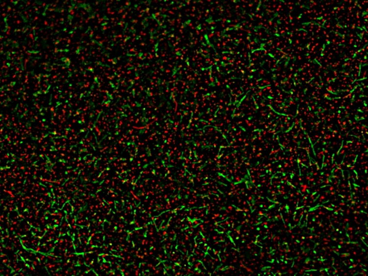
Two-color discrimination of live and dead Bacillus subtilis populations using the MycoLight™ Fluorescence Live/Dead Bacterial Imaging Kit (Cat No. 22411). Live bacteria with active intracellular esterase were labeled with MycoLight™ 520 (green), while 70% alcohol-killed dead bacteria with compromised membranes were labeled with propidium iodide (red).
Neither dye is fixable with glutaraldehyde or paraformaldehyde (PFA). However, they can be used in fixed and permeabilized cells for cell cycle analysis by DNA content measurement (i.e., PI will require the addition of RNase). Alternatively, the high affinity of 7-AAD for GC-rich regions of DNA makes it useful for chromosome banding studies. The binding of 7-AAD to DNA yields a distinct pattern in polytene chromosomes and chromatin.
Dimeric Cyanine Dyes
The dimeric cyanine dye series of DiTO™ and DiYO™ also display exceptional sensitivity for various nucleic acid species, including double-stranded DNA (dsDNA), single-stranded DNA (ssDNA), and RNA. They exhibit a 100 to 1000-fold fluorescence enhancement upon nucleic acid binding, and their molar extinction coefficients are an order magnitude greater than ethidium homodimers. DiYO™-1 and DiTO™-1 dyes are optimally excited by the 488 nm and 514 nm spectral line of the argon-ion laser, respectively, and emit green fluorescence. DiYO™-3 and DiTO™-3 dyes are well-excited by the He-Ne laser and emit red fluorescence. All of the dyes in the DiYO™ and DiTO™ series are supplied as 5 mM solutions in DMSO, except for DiYO™-1, which is also available as a solid. Cyanine monomer dyes TWO-PRO™ 1 and TWO-PRO™ 3 may also be used to analyze DNA content in dead cells.
Table 3. Specifications for PI, 7-AAD, and cyanine dimer dyes.
| Product ▲ ▼ | Ex (nm) ▲ ▼ | Em (nm) ▲ ▼ | ε (cm-1M-1) ▲ ▼ | Φ ▲ ▼ | Spectrum ▲ ▼ | Unit Size ▲ ▼ | Cat No. ▲ ▼ |
| Propidium Iodide | 537 | 618 | 6,000 | 0.2 | 25 mg | 17515 | |
| Propidium Iodide | 537 | 618 | 6,000 | 0.2 | 5g | 17516 | |
| Propidium Iodide *10 mM aqueous solution* | 537 | 618 | 6,000 | 0.2 | 1 mL | 17517 | |
| 7-AAD [7-Aminoactinomycin D] | 549 | 648 | 27,500 | 0.02 | 1 mg | 17501 | |
| DiTO™-1 [equivalent to TOTO®-1] *5 mM DMSO solution* | 514 | 531 | 117,000 | 0.34 | 0.2 mL | 17575 | |
| DiTO™-3 [equivalent to TOTO®-3] *5 mM DMSO solution* | 642 | 661 | 154,000 | 0.06 | 0.2 mL | 17576 | |
| DiYO™-1 [equivalent to YOYO®-1] | 491 | 508 | 98,900 | 0.52 | 1 mg | 17579 | |
| DiYO™-1 [equivalent to YOYO®-1] *5 mM DMSO solution* | 491 | 508 | 98,900 | 0.52 | 0.2 mL | 17580 | |
| DiYO™-3 [equivalent to YOYO®-3] *5 mM DMSO solution* | 612 | 631 | 167,000 | 0.15 | 0.2 mL | 17581 | |
| TWO-PRO™ 1 [equivalent to TO-PRO®-1] *5 mM DMSO solution* | 515 | 531 | 62,800 | 0.25 | 0.2 mL | 17571 |
Nuclear DCS1™ Dead Cell Stains
Nuclear DCS1™ dead cell stains are a family of membrane-impermeant, high-affinity nucleic acid dyes for analyzing DNA content in dead, fixed, or permeabilized cells. These dyes easily penetrate the compromised membranes of dead cells and selectively bind to DNA, producing exceptionally bright signals. In aqueous media, Nuclear DCS1™ dyes are essentially non-fluorescent and can be conveniently added to cells without an additional wash step and imaged with minimal interference. The fluorescence absorption and emission spectra of Nuclear DCS1™ dyes span the visible-light spectrum from violet to red, making them compatible with various fluorescence platforms, including fluorescence microscopes, flow cytometers, and microplate readers. Moreover, the multiple single-color formats of these dyes provide the necessary flexibility to facilitate multicolor applications in imaging and flow cytometry. All dyes in the Nuclear DCS1™ series are supplied as 5 mM solutions in DMSO.
Amine-Reactive Cell Meter™ Fixable Viability Dyes
Cell Meter™ fixable viability dyes are membrane-impermeant, amine-reactive dyes designed to discriminate live and dead cell populations in flow cytometry. These dyes readily penetrate dead cells with compromised membrane integrity and bind irreversibly to cellular amine proteins and other intracellular components producing significant cellular fluorescence. In live cells, these dyes cannot penetrate the membrane leaving only cell-surface amines to bind with and dimly fluoresce. The difference in intensity between viable and dead cells is significant, making it easy to exclude dead cells from analysis. Because staining is robust and stable, cells labeled with Cell Meter™ fixable viability dyes can be fixed, permeabilized, and probed for intracellular immunophenotyping. The Cell Meter™ fixable viability dyes are available in 9 fluorescent stains to provide users greater flexibility in multicolor panel design. Dyes included in this series are excited by primary excitation sources, including the 405, 488, 633, and 647 nm laser lines, and emit in common channels.
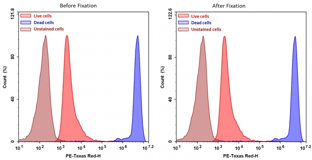
Detection of Jurkat cell viability using Cell Meter™ BX590 fixable viability dye. Jurkat cells were treated and stained with Cell Meter™ BX590 (Cat No. 22514), fixed in 3.7% formaldehyde, and analyzed by flow cytometry. The dead cell population (blue peak) is easily distinguished from the live cell population (red peak) with the PE-Texas Red channel. Nearly identical results were obtained before and after fixation.
Table 5. Fixable viability dyes for live/dead cell analysis during flow cytometry
| Product ▲ ▼ | Ex (nm) ▲ ▼ | Em (nm) ▲ ▼ | Spectrum ▲ ▼ | Unit Size ▲ ▼ | Cat No. ▲ ▼ |
| Cell Meter™ VX450 fixable viability dye | 406 | 445 | 200 Tests | 22540 | |
| Cell Meter™ VX500 fixable viability dye | 433 | 498 | 200 Tests | 22542 | |
| Cell Meter™ BX520 fixable viability dye | 491 | 516 | 200 Tests | 22510 | |
| Cell Meter™ BX590 fixable viability dye | 492 | 579 | 200 Tests | 22514 | |
| Cell Meter™ BX650 fixable viability dye | 518 | 654 | 200 Tests | 22520 | |
| Cell Meter™ RX660 fixable viability dye | 649 | 664 | 200 Tests | 22530 | |
| Cell Meter™ RX700 fixable viability dye | 690 | 713 | 200 Tests | 22532 | |
| Cell Meter™ RX780 fixable viability dye | 629 | 767 | 200 Tests | 22536 | |
| Cell Meter™ IX830 fixable viability dye | 811 | 822 | 200 Tests | 22529 | |
| ReadiView™ Green/Red Viability Stain | 100 Tests | 22762 |
Measuring Enzymatic Activity in Live Cells
Live cells contain several non-specific cytosolic esterases responsible for catalyzing ester hydrolysis, a critical reaction in cell and drug metabolism. The activity of these enzymes can be detected with a wide variety of fluorogenic esterase substrates, including calcein AM, BCECF AM, and carboxyfluorescein diacetate (CFDA), and used to identify live cell populations. As near-neutral compounds, esterase substrates diffuse freely and noninvasively across the plasma membrane and permeate the cytosol of live cells. Once inside the cell, cytosolic esterases rapidly hydrolyze these non-fluorescent substrates into highly fluorescent polar products retained by cells with intact membrane integrity. Labeled cells can be measured by fluorescence microscopy, flow cytometry, or a fluorescence microplate reader. They can be combined with apoptotic and necrotic indicators for multiparameter cell health analysis.
Calcein AM Dyes for Labeling Live Cells
Calcein AM (i.e., a calcein dye synthesized with acetoxymethyl esters (AM)) is a cell-permeable, fluorogenic esterase substrate used to identify live cells. Modifying calcein and other polar compounds with AM groups promotes cell permeability by increasing their hydrophobicity and, more importantly, makes esterase hydrolyzation a requirement to activate their fluorescence. Following brief incubation and hydrolysis of calcein-AM, viable cells will fluoresce bright green when excited by the 488 nm spectral line of the argon-ion laser or with any other blue light excitation source. Compared to other esterase substrates, such as BCECF AM or CFDA, the brighter fluorescence, photostability, and low cytotoxicity of calcein-AM are advantageous for various studies, including cell tracking, cell adhesion, chemotaxis, multidrug resistance, cell viability, apoptosis, and cytotoxicity.
Consequently, the fluorescence emission maximum of calcein at ~521 nm makes it challenging to use in combination with GFP-transfected cells or other green fluorescent indicators for multiplex analysis. To address the single color limitation of calcein-AM, AAT Bioquest has developed calcein derivatives in a broad range of color options, including Calcein Blue AM, Calcein Orange™ AM, and Calcein Red™ AM, and Calcein Deep Red™ AM dyes for multicolor labeling and functional analysis of live cells. For instance, calcein Blue AM is a UV-light excitable viability indicator that emits fluorescence maximally at ~441 nm and is designed for use with instruments optimized for detecting blue fluorescence. It can be combined with the phosphatidylserine stain Annexin V iFluor® 488 (Cat No. 20071) and nucleic acid dead cell stain 7-AAD for the three-color discrimination of live/apoptotic/necrotic cell populations.
CytoCalcein™ Violet Viability Dyes Optimized for Flow Cytometry
CytoCalcein™ Violet 450 and CytoCalcein™ Violet 500 are optimized for use in flow cytometry. The enzymatic conversion of non-fluorescent CytoCalcein™ Violet 450 AM and CytoCalcein™ Violet 500 AM to highly fluorescent CytoCalcein™ Violet 450 and CytoCalcein™ Violet 500 products (Ex/Em maxima ~406/445 nm and ~420/505 nm, respectively) are efficiently excited by the standard 405 nm violet laser found in most flow cytometers. In conjunction with red fluorescent nucleic acid dead cell stains propidium iodide (PI, Ex/Em maxima ~537/618 nm) or 7-Aminoactinomycin D (7-AAD, Ex/Em ~549/648 nm), CytoCalcein™ Violet dyes are excellent fluorophores for two-color discrimination of live from dead cell populations based on membrane integrity and esterase activity. Removing data points representing dead cells is a critical step in improving the accuracy of your flow cytometry analysis. Often dead cells will bind non-specifically to many reagents contributing to false-positive results.
Table 6. Specifications for calcein AM and calcein AM derivatives.
| Product ▲ ▼ | Ex (nm) ▲ ▼ | Em (nm) ▲ ▼ | Spectrum ▲ ▼ | Unit Size ▲ ▼ | Cat No. ▲ ▼ |
| Calcein Blue, AM *CAS 168482-84-6* | 354 nm | 441 nm | 1 mg | 22007 | |
| Calcein UltraBlue™ | 359 nm | 458 nm | 1 mg | 21908 | |
| CytoCalcein™ Violet 450 *Excited at 405 nm* | 406 nm | 445 nm | 1 mg | 22012 | |
| CytoCalcein™ Violet 500 *Excited at 405 nm* | 420 nm | 505 nm | 1 mg | 22013 | |
| Calcein, AM *CAS 148504-34-1* | 501 nm | 521 nm | 1 mg | 22002 | |
| Calcein, AM *UltraPure grade* *CAS 148504-34-1* | 501 nm | 521 nm | 1 mg | 22003 | |
| Calcein, AM *UltraPure grade* *CAS 148504-34-1* | 501 nm | 521 nm | 20x50 µg | 22004 | |
| Calcein Orange™ diacetate | 531 nm | 545 nm | 1 mg | 22009 | |
| Calcein Red™ AM | 562 nm | 576 nm | 1 mg | 21900 | |
| Calcein Deep Red™ AM ester | 643 nm | 663 nm | 1 mg | 22011 |
Measuring Metabolic Activity in Live Cells

Cell proliferation was determined using Cell Meter™ Colorimetric MTT Cell Proliferation Kit. HeLa cells at 0 to 40,000 cells/well/100 µL were added to a clear bottom 96-well plate overnight. The absorbance was measured at 560 nm using a SpectraMax reader (Molecular Devices). 500 cells/well was detected compared to ~5,000 cells/well with Sigma's MTT assay kit.
Tetrazolium Salts for Cell Viability
The four tetrazolium salts used to measure viability are MTT, MTS, XTT, and WST-8. Of the four, MTT is positively charged and can readily penetrate viable cells. The others (MTS, XTT, and WST-8) are negatively charged and membrane-impermeant and require an intermediate electron acceptor that can transfer electrons from the cytoplasm or plasma membrane to facilitate reduction into colored formazan products. Reduction of MTT is the most common. However, for most commercial MTT assays, solubilization of the purple formazan product in DMSO or isopropanol is required before quantification. The convenient Cell Meter™ Colorimetric MTT Cell Proliferation Kit (Cat No. 22768) simplifies the task of counting cells to an easy mix and read format without the need for solubilization. The concentration of formazan determined by optical density at 560 nm is directly proportional to the number of live cells.
Among the other tetrazolium salts, the water-soluble WST-8 offers the highest sensitivity. Reduction of WST-8 by cellular dehydrogenases yields water-soluble orange formazan crystals that do not require solubilization before quantification. The concentration of formazan product can be determined by measuring absorbance at 460 nm and is proportional to the number of living cells. The Cell Meter™ Colorimetric WST-8 Cell Quantification Kit provides a sensitive assay with exceptional linearity of up to ~106 cells per well. No pre-mixing of components is required, as the WST-8 substrate is conveniently provided in a solution that can be directly added to the cells. Each kit provides sufficient reagents for either ~1000 (22770) or ~5000 tests (22771) using a standard 96-well microplate. WST-8 is also available as a stand-alone reagent in 25 mg (Cat No. 15705), 100 mg (Cat No. 15706), and 1 g (Cat No. 15707) unit sizes supplied as 50 mM aqueous solutions.
Table 7. Colorimetric MTT and WST-8 tetrazolium salt-based assays for measuring cell proliferation.
| Product ▲ ▼ | Unit Size ▲ ▼ | Cat No. ▲ ▼ |
| Cell Meter™ Colorimetric MTT Cell Proliferation Kit | 1000 Tests | 22768 |
| Cell Meter™ Colorimetric MTT Cell Proliferation Kit | 5000 Tests | 22769 |
| Cell Meter™ Colorimetric WST-8 Cell Quantification Kit | 1000 Tests | 22770 |
| Cell Meter™ Colorimetric WST-8 Cell Quantification Kit | 5000 Tests | 22771 |
| Cell Counting Kit 8 (CCK-8) | 1000 Tests | 22772 |
| Cell Counting Kit 8 (CCK-8) | 10000 Tests | 22773 |
| ReadiUse™ WST-8 *50 mM aqueous solution* | 25 mg | 15705 |
| ReadiUse™ WST-8 *50 mM aqueous solution* | 100 mg | 15706 |
| ReadiUse™ WST-8 *50 mM aqueous solution* | 1 g | 15707 |
| ReadiUse™ MTS Reagent *50 mM aqueous solution* | 5 mL | 15710 |
Resazurin
Resazurin, also known as Alamar Blue, is a water-soluble redox-sensitive indicator that can be used to measure the cell viability of mammalian cells, bacteria, and fungi. In live metabolically active cells, the blue-colored and non-fluorescent resazurin is irreversibly reduced to a pink-colored and highly-fluorescent resorufin by aerobic respiration. The signal output may be measured using a standard spectrophotometer at 570 nm or by fluorimetry using a 544/590 nm filter set and is proportional to the number of viable cells over a wide concentration range. Since resazurin is minimally toxic to live cells, it is helpful for quantitatively measuring cell proliferation and other long-term studies.
Table 8. Product specifications for resazurin.
| Product ▲ ▼ | Abs (nm) ▲ ▼ | Ex/Em (nm) ▲ ▼ | Unit Size ▲ ▼ | Cat No. ▲ ▼ |
| Resazurin, sodium salt *CAS 62758-13-8* | 605 | 571/584 | 100 mg | 15700 |
Table 9. Yeast/Fungi Cell Viabilty Dyes
| Cat No. ▲ ▼ | Product ▲ ▼ | Unit Size ▲ ▼ |
| 22470 | FunXite™-1 *10 mM DMSO solution* | 100 uL |


