Calcein
Deep Red fluorescence
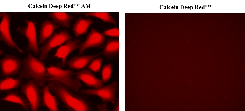
Calcein AM is one the most popular fluorescent probes used for labeling and monitoring cellular functions of live cells. However, the single color of Calcein AM makes it impossible to use this valuable reagent in the multicolor applications. For example, it is impossible to use Calcein AM in GFP-tranfacted cells due to the same color to GFP. To address this color limitation of Calcein AM, we have developed Calcein Orange™, Calcein Red™ and Calcein Deep Red™. These new Calcein AM analogs enable the multicolor labeling and functional analysis of live cells in combination with Calcein AM. Calcein Deep Red™ is the esterase-hydrolyzed product of Calcein Deep Red™ acetate in cells. Calcein Deep Red™ dye can be monitored with the common Cy5 filter set. It is used as a fluorescence reference standard for Calcein Deep Red™ acetate.
Example protocol
Calculators
Common stock solution preparation
Table 1. Volume of DMSO needed to reconstitute specific mass of Calcein Deep Red™ to given concentration. Note that volume is only for preparing stock solution. Refer to sample experimental protocol for appropriate experimental/physiological buffers.
| 0.1 mg | 0.5 mg | 1 mg | 5 mg | 10 mg | |
| 1 mM | 267.623 µL | 1.338 mL | 2.676 mL | 13.381 mL | 26.762 mL |
| 5 mM | 53.525 µL | 267.623 µL | 535.246 µL | 2.676 mL | 5.352 mL |
| 10 mM | 26.762 µL | 133.811 µL | 267.623 µL | 1.338 mL | 2.676 mL |
Molarity calculator
Enter any two values (mass, volume, concentration) to calculate the third.
| Mass (Calculate) | Molecular weight | Volume (Calculate) | Concentration (Calculate) | Moles | ||||
| / | = | x | = |
Spectrum
Alternative formats
| Name | Color | Form |
| Calcein *UltraPure Grade* *CAS 207124-64-9* | Green | - |
| Calcein Red™ sodium salt | Red | Sodium Salt |
| Calcein Deep Red™ | Deep Red | - |
| Calcein UltraBlue™ sodium salt | UltraBlue | Sodium Salt |
| Calcein Blue *CAS 54375-47-2* | Blue | - |
| Calcein Orange™ sodium salt | Orange | Sodium Salt |
| Calcein Orange™ diacetate | Orange | Diacetate |
| Calcein Deep Red™ acetate | Deep Red | Acetate |
Product family
| Name | Excitation (nm) | Emission (nm) |
| Calcein Red™ AM | 562 | 576 |
Citations
View all 20 citations: Citation Explorer
Maternal milk cell components are uptaken by infant liver macrophages via extracellular vesicle mediated transport
Authors: Doerfler, Rose and Yerneni, Saigopalakrishna and LoPresti, Samuel and Chaudhary, Namit and Newby, Alexandra and Melamed, Jilian R and Malaney, Angela and Whitehead, Kathryn A
Journal: The FASEB Journal (2025): e70340
Authors: Doerfler, Rose and Yerneni, Saigopalakrishna and LoPresti, Samuel and Chaudhary, Namit and Newby, Alexandra and Melamed, Jilian R and Malaney, Angela and Whitehead, Kathryn A
Journal: The FASEB Journal (2025): e70340
Skin-Targeted Delivery of Extracellular Vesicle-Encapsulated Curcumin Using Dissolvable Microneedle Arrays
Authors: Yerneni, Saigopalakrishna S and Yalcintas, Ezgi P and Smith, Jason D and Averick, Saadyah and Campbell, Phil G and Ozdoganlar, O Burak
Journal: Acta Biomaterialia (2022)
Authors: Yerneni, Saigopalakrishna S and Yalcintas, Ezgi P and Smith, Jason D and Averick, Saadyah and Campbell, Phil G and Ozdoganlar, O Burak
Journal: Acta Biomaterialia (2022)
Radioiodination of extravesicular surface constituents to study the biocorona, cell trafficking and storage stability of extracellular vesicles
Authors: Yerneni, Saigopalakrishna S and Solomon, Talia and Smith, Jason and Campbell, Phil G
Journal: Biochimica et Biophysica Acta (BBA)-General Subjects (2022): 130069
Authors: Yerneni, Saigopalakrishna S and Solomon, Talia and Smith, Jason and Campbell, Phil G
Journal: Biochimica et Biophysica Acta (BBA)-General Subjects (2022): 130069
Functional imaging of neuronal activity of auditory cortex by using Cal-520 in anesthetized and awake mice
Authors: Li, Jingcheng and Zhang, Jianxiong and Wang, Meng and Pan, Junxia and Chen, Xiaowei and Liao, Xiang
Journal: Biomedical Optics Express (2017): 2599--2610
Authors: Li, Jingcheng and Zhang, Jianxiong and Wang, Meng and Pan, Junxia and Chen, Xiaowei and Liao, Xiang
Journal: Biomedical Optics Express (2017): 2599--2610
NINJ2--A novel regulator of endothelial inflammation and activation
Authors: Wang, Jingjing and Fa, Jingjing and Wang, Pengyun and Jia, Xinzhen and Peng, Huixin and Chen, Jing and Wang, Yifan and Wang, Chenhui and Chen, Qiuyun and Tu, Xin and others, undefined
Journal: Cellular Signalling (2017)
Authors: Wang, Jingjing and Fa, Jingjing and Wang, Pengyun and Jia, Xinzhen and Peng, Huixin and Chen, Jing and Wang, Yifan and Wang, Chenhui and Chen, Qiuyun and Tu, Xin and others, undefined
Journal: Cellular Signalling (2017)
References
View all 84 references: Citation Explorer
Functional evidence that the self-renewal gene NANOG regulates esophageal squamous cancer development
Authors: Li, Deng and Xiang, Xiaocong and Yang, Fei and Xiao, Dongqin and Liu, Kang and Chen, Zhu and Zhang, Ruolan and Feng, Gang
Journal: Biochemical and Biophysical Research Communications (2017)
Authors: Li, Deng and Xiang, Xiaocong and Yang, Fei and Xiao, Dongqin and Liu, Kang and Chen, Zhu and Zhang, Ruolan and Feng, Gang
Journal: Biochemical and Biophysical Research Communications (2017)
Localized functional chemical stimulation of TE 671 cells cultured on nanoporous membrane by calcein and acetylcholine
Authors: Zibek S, Stett A, Koltay P, Hu M, Zengerle R, Nisch W, Stelzle M.
Journal: Biophys J. (2006)
Authors: Zibek S, Stett A, Koltay P, Hu M, Zengerle R, Nisch W, Stelzle M.
Journal: Biophys J. (2006)
A vaccination and challenge model using calcein marked fish
Authors: Klesius PH, Evans JJ, Shoemaker CA, Pasnik DJ.
Journal: Fish Shellfish Immunol (2006): 20
Authors: Klesius PH, Evans JJ, Shoemaker CA, Pasnik DJ.
Journal: Fish Shellfish Immunol (2006): 20
Novel fluorescence assay using calcein-AM for the determination of human erythrocyte viability and aging
Authors: Bratosin D, Mitrofan L, Palii C, Estaquier J, Montreuil J.
Journal: Cytometry A (2005): 78
Authors: Bratosin D, Mitrofan L, Palii C, Estaquier J, Montreuil J.
Journal: Cytometry A (2005): 78
Cytotoxic effects of 100 reference compounds on Hep G2 and HeLa cells and of 60 compounds on ECC-1 and CHO cells. I mechanistic assays on ROS, glutathione depletion and calcein uptake
Authors: Schoonen WG, Westerink WM, de Roos JA, Debiton E.
Journal: Toxicol In Vitro (2005): 505
Authors: Schoonen WG, Westerink WM, de Roos JA, Debiton E.
Journal: Toxicol In Vitro (2005): 505
Page updated on January 23, 2025


