Use TUNEL Assays To Assess DNA Fragmentation
Introduction
Apoptosis or programmed cell death is a highly complex cellular pathway. In response to apoptotic stimuli, cells will undergo a series of controlled steps resulting in the organized destruction, sorting and recycling of vital cellular components. Apoptotic cells can be characterized by distinct morphological and biochemical markers as they progress down the apoptotic pathway. Such features may include cell shrinkage, chromatin condensation, the formation of membrane-bound apoptotic bodies and the expression of phosphatidylserine (PS) phospholipids on the outer leaflet of the plasma membrane. However, one section of this pathway, considered to be a "hallmark" indicator of apoptotic cells is the fragmentation of internucleosomal DNA by activated endonucleases and the horizontal transferring of this genetic material via apoptotic bodies which are phagocytized by neighboring cells.
Apoptotic DNA fragmentation is a result of DNA degradation by the endonuclease DNA fragmentation factor, or DFF. Wang and colleagues (Liu et al. 1997) identified DFF as a heterodimer protein comprised of DNA fragmentation factors 45 and 40 (DFF45 and DFF40). DFF45 behaves as an inhibitor impeding the cleaving functionality of DFF40. Activation of DFF40 is initiated by the cleavage of DFF45 by the effector enzyme caspase 3. Upon dissociation of DFF45, activated DFF40 can initiate cleaving of chromosomal DNA at internucleosomal sites. Cleavage at these sites results in oligonucleosomal DNA fragments approximately 180-200 base pairs in size. Identification of such DNA fragments can be visualized as a ladder-like pattern using agarose gel electrophoresis. However, this technique is laborious, time-consuming and mainly used for gathering qualitative data. For a more sophisticated and well-established method for detecting internucleosomal DNA fragmentation, we recommended utilizing terminal deoxynucleotidyl transferase dUTP nick end labeling or TUNEL assays.
TUNEL Assay
How it works
The TUNEL assay is an in situ DNA staining technique sensitive at detecting DNA strand breaks or DNA fragmentation. It employs a specialized DNA polymerase known as terminal deoxynucleotidyl transferase (TdT) to detect and enzymatically label the free 3' –OH ends of fragmented DNA with dye-modified dUTP nucleotides. With the aid of fluorimetric instruments, TUNEL labeled DNA fragments can be visualized and measured. In contrast to the qualitative agarose gel electrophoresis approach, TUNEL assays provide quantitative detection on apoptosis at a single-cell level (Figure 1).
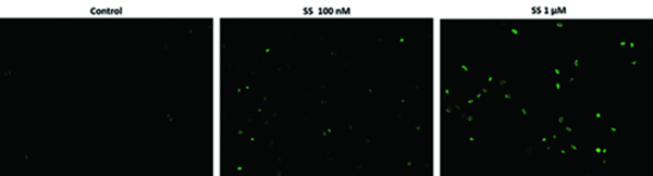
Fluorescence images of TUNEL reaction in HeLa cells with the treatment of 100 nM or 1 µM staurosporin (SS) for 4 hours as compare to untreated control. Cells were incubated with reaction mixture for 1 hour at 37°C. The green fluorescence signal was analyzed using fluorescence microscope with a FITC filter set. Fluorescently labeled DNA strand breaks shows intense fluorescent staining in SS treated cells.
Throughout the years, several variants of the TUNEL technique have been designed and optimized for various chromogenic and fluorogenic detection formats. Chromogenic TUNEL assays are well-suited for us in tissue samples. Whereas, fluorogenic TUNEL assays are designed for use with suspended or adherent cell samples. Several variables influence the staining kinetics of TUNEL assays. Some of these variables include reagent concentration, fixation of sample, and accessibility of DNA strand breaks, which may vary between tissue or cell sample types. Therefore, standardizing your chosen TUNEL assay with positive and negative controls can alleviate these potential influences reducing any chance of detecting a false signal. For a detailed protocol, including positive and negative control steps, please refer to the TUNEL protocol below.
Several kits for the detection of DNA fragmentation are commercially available. With so many possibilities and little variation between competing products, selecting the proper TUNEL assay can prove to be quite difficult. To aid in the selection process we recommend Cell Meter™ TUNEL Apoptosis Assay Kits available in either green or red fluorescence emissions. These green and red TUNEL assays utilize a proprietary dye-modified dUTP nucleotide known as Tunnelyte™ Green and Tunnelyte™ Red, respectively. Tunnelyte™ Green's peak absorption at 497 nm makes it well-suited for efficient excitation by instruments equipped with the 488 nm Argon laser line. While Tunnelyte™ Red's peak absorption at 556 nm is efficiently excited by instruments equipped with the krypton ion laser. Both probes are well separated from DNA staining dyes such as Hoechst (Cat# 17523), DAPI (Cat# 17511) or Nuclear Violet™ LCS1 (Cat#17543), making any an appropriate DNA counterstaining dye.
Compared to other commercially available TUNEL assays which are offered at an average price of $529 with an average unit size of 45 tests, Cell Meter™ TUNEL assays offer the best value with each kit priced at $295 and a unit size of 50 tests. Additionally, the reaction buffers provided in the Cell Meter™ TUNEL kits are free of the extremely hazardous arsenic compound, sodium cacodylate. The highly carcinogenic and toxic arsenic compound make the tunel assay kits from other vendors extremely difficult to be handled, stored and disposed of.
TUNEL Assay Protocol (Sample Type: Adherent or Non-Adherent Cells)
Prepare Cell Culture:
- According to your specific protocol, culture cells to an optimal density for apoptosis induction.
- For adherent cells grown in a 96 well microplate culture, we recommend about 30,000 to 50,000 cells/well.
- For non-adherent cells, we recommend about 1 to 2 x 10⁶ cells/mL.
- At the same time, culture a non-induced negative control cell population at the same specifications as the induced sample for every labeling condition.
Fixation and Permeabilization:
- Remove cell media.
- For adherent cells, add 100 µL/well/96-well plate of 4% formaldehyde fixative buffer to each well.
- Note: For non-adherent cells, add desired amount, such as 2 x 10⁶ cells/mL of 4% formaldehyde fixative buffer.
- Incubate plates for 20 to 30 minutes at room temperature.
- Remove fixative.
Optional step to improve permeability after fixation: Add 100 µL/well/96-well plate of permeabilization reagent, such as 0.2% Triton X-100 in PBS, to each well and incubate the plate for 10 minutes at room temperature.
- Wash the cells with PBS 2 to 3 times.
Optional positive control step: To prepare a positive control for TUNEL reaction, digest cells with DNAse 1 for 30 minutes at room temperature before performing the TUNEL reaction.
TUNEL Reaction:
Note: Prior to beginning TUNEL reaction, each cell line should be evaluated on an individual basis to determine the optimal cell density.
- Prepare reaction mixture based on the number of samples to be assayed:
- Add 0.5 µL of 100X Tunnelyte™ Green to 50 µL of Reaction Buffer for a total volume of 50.5 µL.
- Add 50 µL of freshly prepared reaction mixture (from Step 8.1) to each sample and incubate at 37 °C for 60 minutes.
- Remove reaction mixture and was the cells 3 to 5 times with 200 µL/well of PBS.
- Monitor the fluorescence intensity using a fluorescence microscope, flow cytometer, or fluorescence microplate reader at Ex/Em = 490/520 nm.
Optional: Counterstain the nucleus with 1X Hoechst (Component C, Ex/Em = 350/460 nm) for image analysis
Table 1. Cell Meter Tunel Assays
| Cat No. ▲ ▼ | Product Name ▲ ▼ | Ex (nm) ▲ ▼ | Em (nm) ▲ ▼ |
| 22849 | Cell Meter™ Live Cell TUNEL Apoptosis Assay Kit *Green Fluorescence* | 498 | 522 |
| 22844 | Cell Meter™ Live Cell TUNEL Apoptosis Assay Kit *Red Fluorescence* | 549 | 648 |
References
- Darzynkiewicz, Zbigniew, et al. "Analysis of apoptosis by cytometry using TUNEL assay."Methods, vol. 44, no. 3, 2008, pp. 250–254., doi:10.1016/j.ymeth.2007.11.008.
- Liu, Xuesong, et al. "DFF, a Heterodimeric Protein That Functions Downstream of Caspase-3 to Trigger DNA Fragmentation during Apoptosis."Cell, vol. 89, no. 2, 1997, pp. 175–184., doi:10.1016/s0092-8674(00)80197-x.
- Lu, Z., et al. "Nucleoplasmin regulates chromatin condensation during apoptosis."Proceedings of the National Academy of Sciences, vol. 102, no. 8, July 2005, pp. 2778–2783., doi:10.1073/pnas.0405374102.
- Tang, Damu, and Vincent J. Kidd. "Cleavage of DFF-45/ICAD by Multiple Caspases Is Essential for Its Function during Apoptosis."Journal of Biological Chemistry, vol. 273, no. 44, 1998, pp. 28549–28552., doi:10.1074/jbc.273.44.28549.
Original created on November 30, 2017, last updated on November 8, 2022
Tagged under: