logo
AAT Bioquest

Wheat Germ Agglutinin (WGA)

Wheat germ agglutinin (WGA) is a well-studied lectin known for its binding affinity to N-acetyl-D-glucosamine and sialic acid, making it a valuable tool in various biological applications. Its interaction with glycoconjugates enables widespread use of WGA derivatives and conjugates for fluorescence imaging and analysis, facilitating the labeling of yeast bud scars, fibrotic scar tissue, and the cell membranes of gram bacteria and mammalian cells. WGA specifically targets sequences of β-1,4-GlcNAc-linked residues known as chitodextrins. Each monomer contains two identical, non-interacting binding sites complementary to 3 or 4 β-1,4-GlcNAc units. Among the monosaccharides tested, only GlcNAc shows strong binding to WGA, while ManNAc demonstrates no binding, and GalNAc exhibits weak binding. The mFluor™ Violet 500 labeled WGA is well-excited by the violet laser, emitting a bright green fluorescence at 501 nm. Notably, the mFluor™ Violet 500 WGA conjugate retains its ability to bind to sialic acid and N-acetylglucosaminyl residues, enhancing its utility in fluorescence imaging and analysis of various scientific investigations.
Example protocol

PREPARATION OF STOCK SOLUTIONS

Unless otherwise noted, all unused stock solutions should be divided into single-use aliquots and stored at -20 °C after preparation. Avoid repeated freeze-thaw cycles

mFluor™ Violet 500-Wheat Germ Agglutinin (WGA) Conjugate stock solution (200X)

Add 500 µL of ddH2O into the powder form to make a 2 mg/mL stock solution.

Note: The reconstituted conjugate solution can be stored at 2-8 °C for short-term storage or at -20 °C for long-term storage.

PREPARATION OF WORKING SOLUTION

mFluor™ Violet 500-Wheat Germ Agglutinin (WGA) Conjugate working solution (1X)

Add 5 µL of 200X WGA conjugate solution to 1 mL HHBS Buffer.

Note: The optimized staining concentration may be different with different cell lines. The recommended starting concentration is 5-10 µg/mL for live cells.

SAMPLE EXPERIMENTAL PROTOCOL

Warm the vial to room temperature centrifuge briefly before opening. Staining protocols vary with applications. Appropriate dilution of conjugates should be determined experimentally.

Live Cells Stain
  1. Wash cells twice with a HHBS buffer.
  2. Add 100 µL mFluor™ Violet 500-WGA working solution.

  3. Incubate cells with WGA working solution for 10-30 minutes at 37 °C.
  4. Wash cells twice with HHBS buffer.
  5. Image cells on a fluorescence microscope using Ex/Em = 410/501 nm.

Fixed Cells Stain

WGA conjugates can be also used to stain fixed cells.

  1. Fix cells with 4% Formaldehyde in PBS.

    Note: For fixed cell membrane staining, it is recommended to stain without the permeabilization step. A permeabilization step after fixation can facilitate staining intracellular compartments such as Golgi and Endoplasmic Reticulum (ER) structures.

  2. Add 100 µL mFluor™ Violet 500-WGA working solution.

  3. Incubate cells with WGA working solution for 10-30 minutes at room temperature.
  4. Wash cells twice with HHBS buffer.
  5. Image cells on a fluorescence microscope using Ex/Em = 410/501 nm.

Spectrum

Product family

NameExcitation (nm)Emission (nm)Extinction coefficient (cm -1 M -1)Quantum yieldCorrection Factor (260 nm)Correction Factor (280 nm)
mFluor™ Violet 450-Wheat Germ Agglutinin (WGA) Conjugate4064453500010.8110.3380.078
mFluor™ Violet 540-Wheat Germ Agglutinin (WGA) Conjugate4025351800010.2111.3260.543

References

View all 50 references: Citation Explorer
Pig sperm membrane microdomains contain a highly glycosylated 15-25-kDa wheat germ agglutinin-binding protein.
Authors: Kasekarn, Waraporn and Kanazawa, Takeru and Hori, Kazuki and Tsuchiyama, Tomoyuki and Lian, Xue and Garénaux, Estelle and Kongmanas, Kessiri and Tanphaichitr, Nongnuj and Yasue, Hiroshi and Sato, Chihiro and Kitajima, Ken
Journal: Biochemical and biophysical research communications (2012): 356-62
Measurement of wheat germ agglutinin binding with a fluorescence microscope.
Authors: Model, Michael A and Reese, Jennifer L and Fraizer, Gail C
Journal: Cytometry. Part A : the journal of the International Society for Analytical Cytology (2009): 874-81
Characterization of the wheat germ agglutinin binding to self-assembled monolayers of neoglycoconjugates by AFM and SPR.
Authors: Lienemann, Michael and Paananen, Arja and Boer, Harry and de la Fuente, Jesús M and García, Isabel and Penadés, Soledad and Koivula, Anu
Journal: Glycobiology (2009): 633-43
Wheat germ agglutinin-binding glycoproteins are decreased in Alzheimer's disease cerebrospinal fluid.
Authors: Fodero, L R and Sáez-Valero, J and Barquero, M S and Marcos, A and McLean, C A and Small, D H
Journal: Journal of neurochemistry (2001): 1022-6
Distribution of concanavalin A and wheat germ agglutinin binding sites in the rat peripheral nerve fibres revealed by lectin/glycoprotein-gold histochemistry.
Authors: Dolapchieva, S
Journal: The Histochemical journal (1996): 7-12
Page updated on October 8, 2024

Ordering information

Price
Conjugate
Unit size
Catalog Number
2545025452254542549525500255022550325509255122551525525255302553225539255502555925560255612556225563
Quantity
Add to cart

Additional ordering information

Telephone1-800-990-8053
Fax1-800-609-2943
Emailsales@aatbio.com
InternationalSee distributors
Bulk requestInquire
Custom sizeInquire
Technical SupportContact us
Purchase orderSend to sales@aatbio.com
ShippingStandard overnight for United States, inquire for international
Request quotation

Physical properties

Solvent

Water

Spectral properties

Absorbance (nm)

412

Correction Factor (260 nm)

0.769

Correction Factor (280 nm)

0.365

Extinction coefficient (cm -1 M -1)

250001

Excitation (nm)

410

Emission (nm)

501

Quantum yield

0.811

Storage, safety and handling

H-phraseH303, H313, H333
Hazard symbolXN
Intended useResearch Use Only (RUO)
R-phraseR20, R21, R22

Storage

Freeze (< -15 °C); Minimize light exposure
UNSPSC12171501

Platform

Fluorescence microscope

Excitation410 nm
Emission501 nm
Recommended plateBlack wall, clear bottom
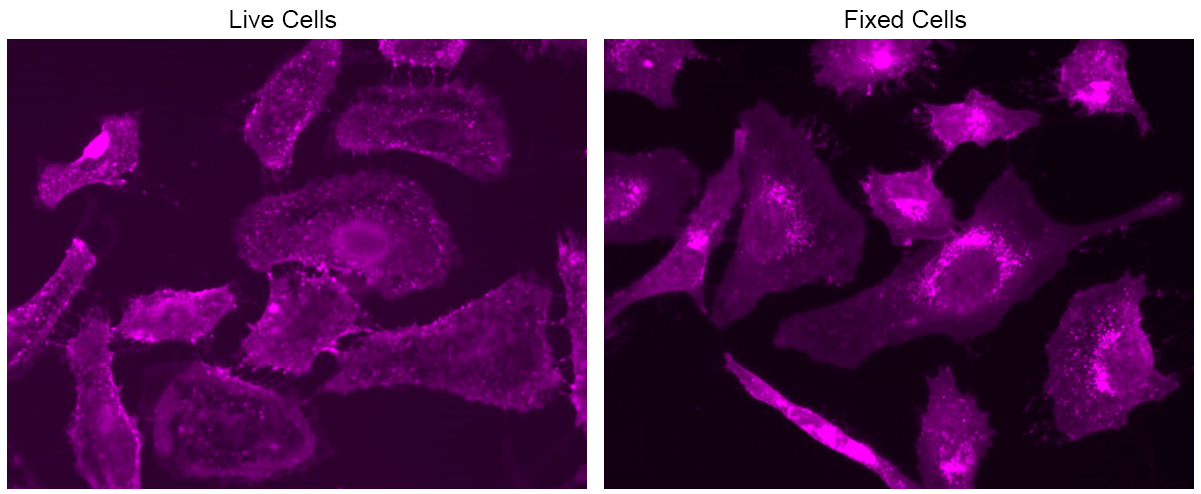
HeLa cells were stained with mFluor™ Violet 500-Wheat Germ Agglutinin (WGA) Conjugate at a concentration of 10 µg/mL for 30 minutes. Images were captured using a fluorescence microscope equipped with a Violet filter set.