logo
AAT Bioquest

Screen Quest™ Fluorimetric Fatty Acid Uptake Assay Kit

Fatty acid uptake is an important therapeutic target for the treatment of many human diseases such as obesity, type 2 diabete and hepatic steatosis. The Screen Quest™ Fluorimetric Fatty Acid Uptake Assay Kit provides a simple and sensitive method for the measurement of fatty acid uptake in cells containing fatty acid transporters. The kit uses a proprietary dodecanoic acid fluorescent fatty acid substrate. This fatty acid uptake assay kit can be performed on any fluorescence microplate reader with a bottom-read mode or FITC channel. The assay can be performed in 96-well or 384-well microtiter plates in a simple mix-and-read procedure, and easily adapted for high throughput screening applications.
Example protocol

AT A GLANCE

Protocol summary

  1. Plate cells in growth medium for 4-6 hours
  2. Transfer the cells into serum free medium for 1 hour and treat cells as desired
  3. Add 100 µL/well of the Fatty Acid dye-loading solution
  4. Monitor fluoroscence increase at Ex/Em = 485/515 nm immediately for kinetics or after 60 minutes incubation for endpoint reading (bottom read mode)

Important notes
Thaw all the kit components at room temperature before starting the experiment.

PREPARATION OF STOCK SOLUTION

Unless otherwise noted, all unused stock solutions should be divided into single-use aliquots and stored at -20 °C after preparation. Avoid repeated freeze-thaw cycles.

TF2-C12 Fatty Acid stock solution:
Add 20 µL of DMSO (Component C) to the vial of TF2-C12 Fatty Acid (Component A) and mix them well. Note:  20 µL of the fluorescent fatty acid substrate stock solution is enough for one plate. The unused fluorescent fatty acid substrate stock solution can be aliquoted and stored at < -20 oC for up to two months if the tubes are sealed tightly and kept from light. Avoid repeated freeze-thaw cycles.

PREPARATION OF WORKING SOLUTION

Fatty Acid dye-loading solution:
Add 20 µL of the TF2-C12 Fatty Acid stock solution to 10 mL of Assay Buffer (Component B) and mix them well. Note: 10 mL of Fatty Acid dye-loading solution is enough for one plate; prepare fresh for each plate and experiment.

SAMPLE EXPERIMENTAL PROTOCOL

Cells preparation:

Prepare cells as desired. The following protocols are guidelines to prepare 3T3-L1adipocytes.

  • Prepare differentiated 3T3-L1 adipocytes (see Ref 1): 3T3-L1 fibroblasts were grown 2 days in a 75 cm flask post-confluence in DMEM/FBS, and then for 2 days in DMEM/FBS supplemented with 0.83 µM insulin, 0.25 µM dexamethasone, and 0.25 mM isobutylmethylxanthine. The medium is changed to maintain the insulin concentration with dexamethasone and IBMX absent for another 2 days. The medium was then changed to DMEM/FBS alone for another 3-5 days. Differentiated cells (at least 95% of which showed an adipocyte phenotype by accumulation of lipid droplets) were used on day 8 to 12 after induction of differentiation.

  • Plate 3T3-L1 adipocytes in growth medium at 50,000-80,000 cells/well/100 µL/96-well or 12,500-20,000 cells/well/25 µL/384-well black wall/clear bottom cell culture Poly-D lysine plate for 4-6 hours before experiment. Centrifuge the plate at 800 rpm for 2 minutes with brake off. Note: It is recommended to plate 3 wells with growth medium only (without cells) as blank wells for data normalization. Note: We find that adipocytes plated at the same day (4-6 hours, and then serum deprived for 1 hour) give better results than that plated for overnight.
  • Remove the cell plate from the incubator, aspirate the medium from the wells, and deprive the cells with 90 µL/well/ 96 well-plate or 20 µL/well/384 well-plate serum free medium. Incubate the cells at 37 ºC, 5% CO2 incubator for 1 hr.

  • Treat the cells by adding 10 µL/well/96-well plate (5 µL/well/384-well plate) of the test compounds or 1X Hanks and 20 mM Hepes buffer (1X HBSS, pH 7.4) or buffer of your choice as the compound diluent.  For blank wells, add the compound diluents. Incubate the cells at 37 ºC, 5% CO2 incubator for a desired period of time (30 minutes for 3T3-L1 cells treated with Insulin).

Sample protocol:

  1. Treat cells with test compounds as desired.

  2. Remove compound-treated cell plates from the incubator, add 100 µL/well (96-well plate) or 25 µL/well (384-well plate) (including blank wells) of the Fatty Acid dye-loading solution.

  3. Measure the fluorescence signal with a fluorescence microplate reader at Ex/Em = 485/515 nm (cut off at 495 nm) using a bottom read mode. Note: For kinetic reading:  Read the fluorescence intensity immediately at 20 seconds interval for 30-60 minutes. Note: For endpoint reading: Read the fluorescence intensity at the end of the 30-60 minutes incubation.

Citations

View all 6 citations: Citation Explorer
Chemically synthesized osteocalcin alleviates NAFLD via the AMPK-FOXO1/BCL6-CD36 pathway
Authors: Zhang, Miao and Dong, Keting and Du, Qian and Xu, Jiaojiao and Bai, Xue and Chen, Lei and Yang, Jianhong
Journal: Journal of Translational Medicine (2024): 1--20
Insulin-stimulated translocation of the fatty acid transporter CD36 to the plasma membrane is mediated by the small GTPase Rac1 in adipocytes
Authors: Chan, Man Piu and Takenaka, Nobuyuki and Abe, Yuki and Satoh, Takaya
Journal: Cellular Signalling (2024): 111102
Mouse Memory CD8 T Cell Subsets Defined by Tissue-Resident Memory Integrin Expression Exhibit Distinct Metabolic Profiles
Authors: Sportiello, Mike and Poindexter, Alexis and Reilly, Emma C and Geber, Adam and Lambert Emo, Kris and Jones, Taylor N and Topham, David J
Journal: ImmunoHorizons (2023): 652--669
PRP19 Enhances Esophageal Squamous Cell Carcinoma Progression by Reprogramming SREBF1-Dependent Fatty Acid Metabolism
Authors: Zhang, Guang-Cong and Yu, Xiang-Nan and Guo, Hong-Ying and Sun, Jia-Lei and Liu, Zhi-Yong and Zhu, Ji-Min and Liu, Tao-Tao and Dong, Ling and Shen, Xi-Zhong and Yin, Jie
Journal: Cancer Research (2023): OF1--OF17
Andrographolide ameliorates hepatic steatosis by suppressing FATP2-mediated fatty acid uptake in mice with nonalcoholic fatty liver disease
Authors: Ran, Li-Sha and Wu, Ya-Zeng and Gan, Yi-Wen and Wang, Hong-Lian and Wu, Li-Juan and Zheng, Chun-Mei and Ming, Yao and Xiong, Ran and Li, Yong-Lin and Lei, Shi-Hang and others,
Journal: Journal of Natural Medicines (2022): 1--14

References

View all 69 references: Citation Explorer
Significance of short chain fatty acid transport by members of the monocarboxylate transporter family (MCT)
Authors: Moschen I, Broer A, Galic S, Lang F, Broer S.
Journal: Neurochem Res (2012): 2562
Improved working memory but no effect on striatal vesicular monoamine transporter type 2 after omega-3 polyunsaturated fatty acid supplementation
Authors: Narendran R, Frankle WG, Mason NS, Muldoon MF, Moghaddam B.
Journal: PLoS One (2012): e46832
Munc18c provides stimulus-selective regulation of GLUT4 but not fatty acid transporter trafficking in skeletal muscle
Authors: Jain SS, Snook LA, Glatz JF, Luiken JJ, Holloway GP, Thurmond DC, Bonen A.
Journal: FEBS Lett (2012): 2428
Emerging risk factors for postpartum depression: serotonin transporter genotype and omega-3 fatty acid status
Authors: Shapiro GD, Fraser WD, Seguin JR.
Journal: Can J Psychiatry (2012): 704
Poultry fat decreased fatty acid transporter protein mRNA expression and affected fatty acid composition in chickens
Authors: Yuan J, Zhang B, Guo Y.
Journal: J Anim Sci Biotechnol (2012): 17
Page updated on January 18, 2025

Ordering information

Price
Unit size
Catalog Number36385
Quantity
Add to cart

Additional ordering information

Telephone1-800-990-8053
Fax1-800-609-2943
Emailsales@aatbio.com
InternationalSee distributors
Bulk requestInquire
Custom sizeInquire
Technical SupportContact us
Purchase orderSend to sales@aatbio.com
ShippingStandard overnight for United States, inquire for international
Request quotation

Storage, safety and handling

H-phraseH303, H313, H333
Hazard symbolXN
Intended useResearch Use Only (RUO)
R-phraseR20, R21, R22
UNSPSC12352200

Platform

Fluorescence microplate reader

Excitation485 nm
Emission515 nm
Cutoff495 nm
Recommended plateBlack wall, clear bottom
Instrument specification(s)Bottom read mode

Components

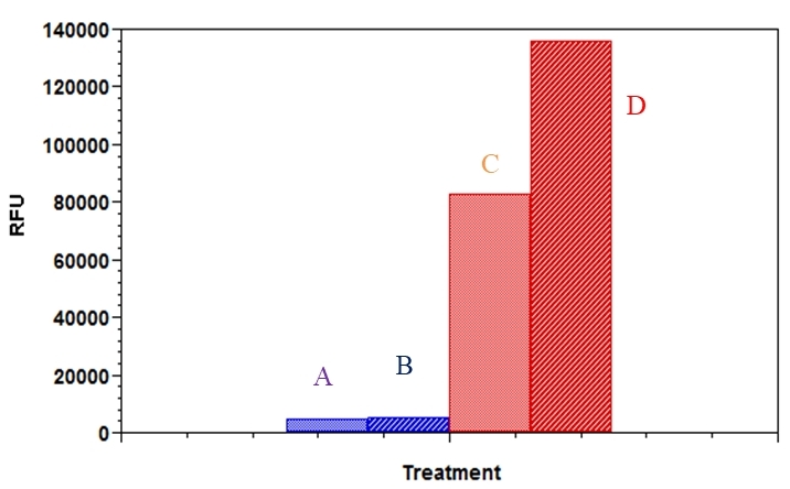
Comparison of fatty acid uptake by 3T3-L1 adipocytes and fibroblast.&nbsp; Cells were plated at 50,000 cells/100 mL/well in a 96 well black wall/clear bottom poly-D lysine plate for 5 hours, and then serum deprived for 1 hour. Cells were treated without (control) or with insulin (150 nM), and incubated at 37 &ordm;C, 5% CO<sub>2 </sub>incubator for 30 min. At the end of the incubation time, 100 &micro;l of fatty acid mixture was added into the well, and incubated for another 60 min, the fluorescence signal was measured with a FlexStation plate reader using bottom read mode. A &ndash; fibroblasts (Control); B &ndash; fibroblasts (Insulin); C &ndash; adipocytes (Control); D&ndash; adipocytes (Insulin).