Portelite™ Fluorimetric RNA Quantitation Kit*Optimized for Cytocite™ and Qubit™ Fluorometers*
Detecting and quantitating small amounts of RNA is extremely important for a wide variety of biological applications such as measuring yields of in vitro transcribed RNA and measuring RNA concentrations before performing Northern blot analysis, S1 nuclease assays, RNase protection assays, cDNA library preparation, reverse transcription PCR, and differential display PCR. The most commonly used technique for measuring nucleic acid concentration is the determination of absorbance at 260 nm. The absorbance-based method is limited by the interferences caused from proteins, free nucleotides and other UV absorbing compounds. The use of sensitive and selective fluorescent nucleic acid stains alleviates this interference problem. StrandBrite™ RNA quantifying reagent is an ultrasensitive fluorescent nucleic acid stain for quantitating RNA in solution. StrandBrite™ RNA quantifying reagent can detect as little as 5 ng/mL RNA with a CytoCite™ or Qubit™ fluorometer. Our StrandBrite™ Green Fluorimetric RNA Quantitation Kit includes our StrandBrite™ Green nucleic acid stain with an optimized and robust protocol. It provides a convenient method for quantifying RNA in solutions.
Example protocol
AT A GLANCE
Protocol summary
- Prepare StrandBrite™ RNA working solution
- Add 190 µL StrandBrite™ RNA working solution into each 0.2 mL PCR tube
- Add 10 µL RNA Standards or test samples into each tube
- Incubate at room temperature for 2 minutes
- Monitor fluorescence with CytoCite™ or Qubit™ fluorometer
Important
Bring all the kit components at room temperature before starting the experiment.PREPARATION OF WORKING SOLUTION
StrandBrite™ RNA working solution
Make a 200-fold dilution of StrandBrite™ Green (Component A) in Assay Buffer (Component B). For example, to prepare enough working solution for 8 samples, add 5 µL of StrandBrite™ Green (Component A) into 1 mL of Assay Buffer (Component B).Note Protect the working solution from light by covering it with foil or placing it in the dark. We recommend preparing this solution in a plastic container rather than glass, as the dye may adsorb to glass surfaces. For best results, this solution should be used within a few hours of its preparation.
SAMPLE EXPERIMENTAL PROTOCOL
The acceptable sample volume could be a range from 1~20 µL depending on the estimate concentration of RNA sample. The recommend sample volume is 10 µL with the RNA concentration in 0.01~10 ng/µL range. If other sample volume is being used, please adjust the dilution factor in the concentration calculations.
The following protocol is generated based on10 µL sample volume with the RNA concentration in 0.01~10 ng/µL range.
- Add 190 µL StrandBrite™ Green working solution into each CytoCite™ sample tube (#CCT100) or equivalent 0.2 mL PCR tube.
Note Use thin-wall, polypropylene, clear 0.2 mL PCR tubes such as#CCT100. - Add 10 µL RNA standard #1 and #2 or test samples into each tube, and then mix by vortexing 2~3 seconds.
- Incubate all tubes at room temperature for 2 minutes.
- Insert the samples into CytoCite™ or Quibit™ and monitor the fluorescence with green fluorescence channel. Follow the procedure appropriate for CytoCite™ Fluorometer. For instructions, see: https://devices.aatbio.com/documentation/user-manual-for-cytocite-fluorometer
PREPARATION OF STANDARD CALIBRATION CURVE (Optional)
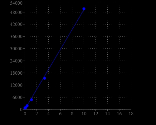
For StrandBrite™ assays, you have the choice to make a calibration curve with the RNA standards. Here is a brief protocol to generate a customized RNA standard curve:- Perform 1/3 serial dilution with Assay Buffer to get 10, 3, 1, 0.3, 0.1, 0.03, 0.01 and 0 ng/µL RNA standard dilutions using RNA Standard #2.
- Add 190 µL StrandBrite™ Green working solution into each tube.
- Add 10 µL RNA standards or test samples into each tube, and then mix by vortexing 2~3 seconds.
- Incubate the reaction at room temperature for 2 minutes.
- Insert the samples into CytoCite™ and monitor the fluorescence with green fluorescence channel.
Spectrum
Open in Advanced Spectrum Viewer
Product family
Name | Excitation (nm) | Emission (nm) |
Portelite™ Fluorimetric ssDNA Quantitation Kit *Optimized for Cytocite™ and Qubit™ Fluorometers* | 498 | 519 |
References
View all 50 references: Citation Explorer
Oncologic Implications of Chronic Hepatitis C Virus Infection.
Authors: Hwang, Jessica P and LoConte, Noelle K and Rice, John P and Foxhall, Lewis E and Sturgis, Erich M and Merrill, Janette K and Torres, Harrys A and Bailey, Howard H
Journal: Journal of oncology practice (2019): 629-637
Authors: Hwang, Jessica P and LoConte, Noelle K and Rice, John P and Foxhall, Lewis E and Sturgis, Erich M and Merrill, Janette K and Torres, Harrys A and Bailey, Howard H
Journal: Journal of oncology practice (2019): 629-637
Outcomes and costs of single-step hepatitis C testing in primary care, Birmingham, United Kingdom.
Authors: Munang, M and Smit, E and Barnett, T and Atherton, C and Tahir, M and Atabani, S F
Journal: Public health (2019): 40-44
Authors: Munang, M and Smit, E and Barnett, T and Atherton, C and Tahir, M and Atabani, S F
Journal: Public health (2019): 40-44
Evaluation of the Aptima HCV Quant Dx Assay Using Serum and Dried Blood Spots.
Authors: Weber, Jenna and Sahoo, Malaya K and Taylor, Nathaniel and Shi, Run-Zhang and Pinsky, Benjamin A
Journal: Journal of clinical microbiology (2019)
Authors: Weber, Jenna and Sahoo, Malaya K and Taylor, Nathaniel and Shi, Run-Zhang and Pinsky, Benjamin A
Journal: Journal of clinical microbiology (2019)
PARP-1/2 Inhibitor Olaparib Prevents or Partially Reverts EMT Induced by TGF-β in NMuMG Cells.
Authors: Schacke, Michelle and Kumar, Janani and Colwell, Nicholas and Hermanson, Kole and Folle, Gustavo A and Nechaev, Sergei and Dhasarathy, Archana and Lafon-Hughes, Laura
Journal: International journal of molecular sciences (2019)
Authors: Schacke, Michelle and Kumar, Janani and Colwell, Nicholas and Hermanson, Kole and Folle, Gustavo A and Nechaev, Sergei and Dhasarathy, Archana and Lafon-Hughes, Laura
Journal: International journal of molecular sciences (2019)
Tumor-draining lymph nodes demonstrate a suppressive immunophenotype in patients with non-small cell lung cancer assessed by endobronchial ultrasound-guided transbronchial needle aspiration: A pilot study.
Authors: Murthy, Vivek and Katzman, Daniel P and Tsay, Jun-Chieh J and Bessich, Jamie L and Michaud, Gaetane C and Rafeq, Samaan and Minehart, Janna and Mangalick, Keshav and de Lafaille, M A Curotto and Goparaju, Chandra and Pass, Harvey and Sterman, Daniel H
Journal: Lung cancer (Amsterdam, Netherlands) (2019): 94-99
Page updated on December 17, 2024