Products
Services
Resources
Selection Guides
About
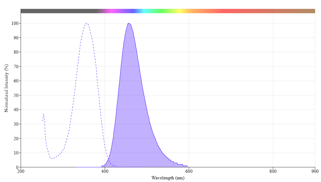
mFluor™ UV460 maleimide
mFluor™ UV460 Maleimide is an excellent building block that can be readily used for labeling biomolecules that have a free thiol (SH) group such as antibodies and thiol-modified oligos. mFluor™ UV460 (MFUV460) dyes are well excited by UV excitation with emission at ~460 nm. MFUV460 has spectral properties almost identical to those of Marina Blue® dye. These spectral characteristics make them an excellent alternative to Marina Blue® dye (Marina Blue® is the trademark of ThermoFisher). AAT Bioquest’s mFluor™ dyes are developed for multicolor flow cytometry-focused applications. These dyes have large Stokes Shifts and can be well excited by the laser lines of flow cytometers (e.g., 355 nm, 405 nm, 488 nm and 633 nm).
| Catalog | Size | Price | Quantity |
|---|---|---|---|
| 1604 | 1 mg | Price |

Physical properties
| Molecular weight | 392.31 |
| Solvent | DMSO |
Spectral properties
| Absorbance (nm) | 364 |
| Correction factor (260 nm) | 0.35 |
| Correction factor (280 nm) | 0.134 |
| Extinction coefficient (cm -1 M -1) | 15000 1 |
| Excitation (nm) | 358 |
| Emission (nm) | 456 |
| Quantum yield | 0.86 1 |
Storage, safety and handling
| H-phrase | H303, H313, H333 |
| Hazard symbol | XN |
| Intended use | Research Use Only (RUO) |
| R-phrase | R20, R21, R22 |
| Storage | Freeze (< -15 °C); Minimize light exposure |
Contact us
| Telephone | |
| Fax | |
| sales@aatbio.com | |
| International | See distributors |
| Bulk request | Inquire |
| Custom size | Inquire |
| Technical Support | Contact us |
| Request quotation | Request |
| Purchase order | Send to sales@aatbio.com |
| Shipping | Standard overnight for United States, inquire for international |
Page updated on June 3, 2026