logo
AAT Bioquest

mFluor™ Blue 660 tyramide

mFluor™ Blue 660 tyramide is a fluorescent labeling reagent used in immunofluorescence staining and in situ hybridization. Tyramide is a small molecule that can diffuse through tissue sections or cell membranes and subsequently be enzymatically amplified to produce a localized, highly fluorescent signal. The mFluor™ Blue 660 dye is conjugated to tyramide to create a fluorescently labeled tyramide that can be used for visualizing specific target molecules or structures within biological samples. mFluor™ Blue 660 tyramide has the largest Stokes Shift among all the commercial tyramide reagents. The high sensitivity and specificity of mFluor™ Blue 660 tyramide make it an excellent choice for fluorescence imaging and detection in various histochemical fluorescence imaging applications.
Example protocol

AT A GLANCE

Protocol Summary
  1. Fix/permeabilize/block cells or tissue
  2. Add primary antibody in blocking buffer
  3. Add HRP-conjugated secondary antibody
  4. Prepare tyramide working solution and apply in cells or tissue for 5-10 minutes at room temperature

PREPARATION OF STOCK SOLUTIONS

Unless otherwise noted, all unused stock solutions should be divided into single-use aliquots and stored at -20 °C after preparation. Avoid repeated freeze-thaw cycles

Tyramide stock solution (200X)

Add 100 µL of DMSO to the vial of mFluor™ tyramide and mix well.

Note: Make single-use aliquots and store unused 200X stock solution at 2-8 °C, protected from light. Avoid repeat freeze-thaw cycles. 

PREPARATION OF WORKING SOLUTION

Tyramide working solution (1X)

Add 100 µL of the tyramide stock solution into 20 mL of a buffer of your choice containing 0.003% H2O2.

Note: For optimal performance, use Tris Buffer, pH=7.4.

Note: A 20 mL solution is good for 200 tests. The tyramide working solution should be used immediately and made fresh on the day of use. Avoid direct exposure to light.

Secondary antibody-HRP working solution

Make an appropriate concentration of secondary antibody-HRP working solution per the manufacturer's recommendations. 

SAMPLE EXPERIMENTAL PROTOCOL

This protocol is applicable for both cells and tissues staining.

Cell fixation and permeabilization
  1. Fix the cells or tissue with 3.7% formaldehyde or paraformaldehyde, in PBS at room temperature for 20 minutes.
  2. Rinse the cells or tissue with PBS twice.
  3. Permeabilize the cells with 0.1% Triton X-100 solution for 1-5 minutes at room temperature.
  4. Rinse the cells or tissue with PBS twice.
Tissue fixation, deparaffinization and rehydration

Deparaffinize and dehydrate the tissue according to the standard IHC protocols. Perform antigen retrieval with the preferred specific solution/protocol as needed. A protocol can be found at:

https://www.aatbio.com/resources/guides/paraffin-embedded-tissue-immunohistochemistry-protocol.html

Peroxidase labeling
  1. Optional: Quench endogenous peroxidase activity by incubating cell or tissue sample in peroxidase quenching solution (such as 3% hydrogen peroxide) for 10 minutes. Rinse with PBS twice at room temperature.
  2. Optional: If using HRP-conjugated streptavidin, it is advisable to block endogenous biotins by biotin blocking buffer.
  3. Block with preferred blocking solution (such as PBS with 1% BSA) for 30 minutes at 4 °C.
  4. Remove blocking solution and add primary antibody diluted in recommended antibody diluent for 60 minutes at room temperature or overnight at 4 °C.
  5. Wash with PBS three times for 5 minutes each.
  6. Apply 100 µL of secondary antibody-HRP working solution to each sample and incubate for 60 minutes at room temperature.

    Note: Incubation time and concentration can be varied depending on the signal intensity.

  7. Wash with PBS three times for 5 minutes each.
Tyramide labeling
  1. Prepare and apply 100 µL of Tyramide working solution to each sample and incubate for 5-10 minutes at room temperature.

    Note: If you observe a non-specific signal, you can shorten the incubation time with the tyramide reagent. You should optimize the incubation period using positive and negative control samples at various incubation time points. Or you can use a lower concentration of the tyramide reagent in the working solution.

  2. Rinse with PBS three times.
Counterstain and fluorescence imaging
  1. Counterstain the cell or tissue samples as needed. AAT provides a series of nucleus counterstain reagents as listed in Table 1. Follow the instruction provided with the reagents.
  2. Mount the coverslip using a mounting medium with anti-fading properties.

    Note: To ensure optimal results, it is recommended to use either ReadiUse™ microscope mounting solution (Cat. 20009) or FluoroQuest™ TSA/PSA Antifade Mounting Medium *Optimized for Tyramide and Styramide Imaging* (Cat. 44890) instead of Vectashield® mounting media. There are instances where Vectashield® mounting media may not be suitable for certain TSA/PSA conjugates.

  3. Use the appropriate filter set to visualize the signal from the Tyramide labeling.

Table 1. Products recommended for nucleus counterstain

Cat#Product NameEx/Em (nm)
17548Nuclear Blue™ DCS1350/461
17550Nuclear Green™ DCS1503/526
17551Nuclear Orange™ DCS1528/576
17552Nuclear Red™ DCS1642/660

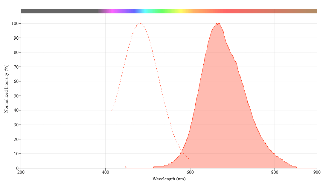
Spectrum

Product family

NameExcitation (nm)Emission (nm)Extinction coefficient (cm -1 M -1)Correction Factor (260 nm)Correction Factor (280 nm)
mFluor™ Blue 660 SE4816632600010.3380.32
mFluor™ Blue 660 Styramide4816632600010.3380.32

References

View all 6 references: Citation Explorer
Use of tyramide-fluorescence in situ hybridization and chromosome microdissection for ascertaining homology relationships and chromosome linkage group associations in oats.
Authors: Sanz, M J and Loarce, Y and Ferrer, E and Fominaya, A
Journal: Cytogenetic and genome research (2012): 145-56
Assignment of N-acetyl-D-glucosaminidase (Mgea5) to rat chromosome 1q5 by tyramide fluorescence in situ hybridization (T-FISH): synteny between rat, mouse and human with Insulin Degradation Enzyme (IDE).
Authors: Van Tine, B A and Patterson, A J and Kudlow, J E
Journal: Cytogenetic and genome research (2003): 202B
Rapid detection and enumeration of Naegleria fowleri in surface waters by solid-phase cytometry.
Authors: Pougnard, Claire and Catala, Philippe and Drocourt, Jean-Louis and Legastelois, Stephane and Pernin, Pierre and Pringuez, Emmanuelle and Lebaron, Philippe
Journal: Applied and environmental microbiology (2002): 3102-7
Tissue distribution of products of the mouse decay-accelerating factor (DAF) genes. Exploitation of a Daf1 knock-out mouse and site-specific monoclonal antibodies.
Authors: Lin, F and Fukuoka, Y and Spicer, A and Ohta, R and Okada, N and Harris, C L and Emancipator, S N and Medof, M E
Journal: Immunology (2001): 215-25
Assignment of human MFNG, manic fringe Drosophila homolog, to 22q13.1 using tyramide fluorescence in situ hybridization (T-FISH).
Authors: Van Tine, B A and Knops, J and Shaw, G M and May, W A
Journal: Cytogenetics and cell genetics (1999): 132-3
Page updated on November 20, 2024

Ordering information

Price
Unit size
Catalog Number11051
Quantity
Add to cart

Additional ordering information

Telephone1-800-990-8053
Fax1-800-609-2943
Emailsales@aatbio.com
InternationalSee distributors
Bulk requestInquire
Custom sizeInquire
Technical SupportContact us
Purchase orderSend to sales@aatbio.com
ShippingStandard overnight for United States, inquire for international
Request quotation

Physical properties

Molecular weight

770.00

Solvent

DMSO

Spectral properties

Absorbance (nm)

481

Correction Factor (260 nm)

0.338

Correction Factor (280 nm)

0.32

Extinction coefficient (cm -1 M -1)

260001

Excitation (nm)

481

Emission (nm)

663

Storage, safety and handling

H-phraseH303, H313, H333
Hazard symbolXN
Intended useResearch Use Only (RUO)
R-phraseR20, R21, R22

Storage

Freeze (< -15 °C); Minimize light exposure
UNSPSC12171501

Platform

Fluorescence microscope

ExcitationViolet filter set
EmissionViolet filter set
Recommended plateBlack wall, clear bottom
Instrument specification(s)Compatible with Cy5 filter set