logo
AAT Bioquest

mFluor™ Blue 659 SE

The mFluor™ Blue 659 dye is optimally excited with a 488 nm blue laser, offering a significant Stokes shift with an emission peak at approximately 659 nm. This water-soluble dye and its protein conjugates exhibit robust red fluorescence when excited at 488 nm, making them ideal for flow cytometry. Compared to RPE (R-Phycoerythrin), mFluor™ Blue 659 dyes are more photostable, enhancing their suitability for fluorescence imaging. Additionally, mFluor™ Blue 659 serves as a distinctive fluorochrome for spectral flow cytometry due to its unique spectral profile, a rarity among available dyes.

The succinimidyl ester (SE) of mFluor™ Blue 659 is a widely used reagent for the conjugation of this dye to proteins or antibodies. Succinimidyl esters react selectively and efficiently with primary amines (such as the side chains of lysine residues or aminosilane-coated surfaces) at pH 7-9, forming stable covalent amide bonds. This property makes mFluor™ Blue 659 SE an excellent choice for labeling proteins, amine-modified oligonucleotides, and other amine-containing molecules.
Example protocol

PREPARATION OF STOCK SOLUTIONS

Unless otherwise noted, all unused stock solutions should be divided into single-use aliquots and stored at -20 °C after preparation. Avoid repeated freeze-thaw cycles

Protein Stock Solution (Solution A)
  1. Mix 100 µL of a reaction buffer (e.g., 1 M  sodium carbonate solution or 1 M phosphate buffer with pH ~9.0) with 900 µL of the target protein solution (e.g., antibody, protein concentration >2 mg/mL if possible) to give 1 mL protein labeling stock solution.

    Note: The pH of the protein solution (Solution A) should be 8.5 ± 0.5. If the pH of the protein solution is lower than 8.0, adjust the pH to the range of 8.0-9.0 using 1 M  sodium bicarbonate solution or 1 M pH 9.0 phosphate buffer.

    Note: The protein should be dissolved in 1X phosphate-buffered saline (PBS), pH 7.2-7.4. If the protein is dissolved in Tris or glycine buffer, it must be dialyzed against 1X PBS, pH 7.2-7.4, to remove free amines or ammonium salts (such as ammonium sulfate and ammonium acetate) that are widely used for protein precipitation.

    Note: Impure antibodies or antibodies stabilized with bovine serum albumin (BSA) or gelatin will not be labeled well. The presence of sodium azide or thimerosal might also interfere with the conjugation reaction. Sodium azide or thimerosal can be removed by dialysis or spin column for optimal labeling results.

    Note: The conjugation efficiency is significantly reduced if the protein concentration is less than 2 mg/mL. The final protein concentration range of 2-10 mg/mL is recommended for optimal labeling efficiency.

mFluor™ Blue 659 SE Stock Solution (Solution B)
  1. Add anhydrous DMSO into the vial of mFluor™ Blue 659 SE to make a 10 mM stock solution. Mix well by pipetting or vortex.

    Note: Before starting the conjugation, prepare the dye stock solution (Solution B). Use promptly. Extended storage of the dye stock solution may reduce the dye activity. Solution B can be stored in the freezer for two weeks when protected from light and moisture. Avoid freeze-thaw cycles.

SAMPLE EXPERIMENTAL PROTOCOL

This labeling protocol was developed for the conjugate of Goat anti-mouse IgG with mFluor™ Blue 659 SE. You might need further optimization for your particular proteins.

Note: Each protein requires distinct dye/protein ratio, which also depends on the properties of dyes. Over labeling of a protein could detrimentally affects its binding affinity while the protein conjugates of low dye/protein ratio gives reduced sensitivity.

Run Conjugation Reaction
  1. Use a 10:1 molar ratio of Solution B (dye)/Solution A (protein) as the starting point:  Add 5 µL of the dye stock solution (Solution B, assuming the dye stock solution is 10 mM) into the vial of the protein solution (95 µL of Solution A) with effective shaking. The concentration of the protein is ~0.05 mM assuming the protein concentration is 10 mg/mL, and the molecular weight of the protein is ~200KD.

    Note: We recommend using a 10:1 molar ratio of Solution B (dye)/Solution A (protein). If it is too low or too high, determine the optimal dye/protein ratio at 5:1, 15:1, and 20:1, respectively.

  2. Continue to rotate or shake the reaction mixture at room temperature for 30-60 minutes.
Purify the Conjugate

The following protocol is an example of dye-protein conjugate purification by using a Sephadex G-25 column.

  1. Prepare Sephadex G-25 column according to the manufacture instruction.
  2. Load the reaction mixture (From "Run conjugation reaction") to the top of the Sephadex G-25 column.
  3. Add PBS (pH 7.2-7.4) as soon as the sample runs just below the top resin surface.
  4. Add more PBS (pH 7.2-7.4) to the desired sample to complete the column purification. Combine the fractions that contain the desired dye-protein conjugate.

    Note: For immediate use, the dye-protein conjugate need be diluted with staining buffer, and aliquoted for multiple uses.

    Note: For longer term storage, dye-protein conjugate solution need be concentrated or freeze dried.

Characterize the Desired Dye-Protein Conjugate

The Degree of Substitution (DOS) is the most important factor for characterizing dye-labeled protein. Proteins of lower DOS usually have weaker fluorescence intensity, but proteins of higher DOS (e.g., DOS > 6) tend to have reduced fluorescence too. The optimal DOS for most antibodies is recommended between 2 and 10, depending on the properties of dye and protein. For effective labeling, the degree of substitution should be controlled to have 6-8 moles of mFluor™ Blue 659 SE to one mole of antibody. The following steps are used to determine the DOS of mFluor™ Blue 659-labeled proteins.

Measure Absorption

To measure the absorption spectrum of a dye-protein conjugate, it is recommended to keep the sample concentration in the range of 1-10 µM depending on the extinction coefficient of the dye.

Read OD (absorbance) at 280 nm and dye maximum absorption (ƛmax = 659 nm for mFluor™ Blue 659 dyes

For most spectrophotometers, the sample (from the column fractions) needs to be diluted with de-ionized water so that the O.D. values are in the range of 0.1 to 0.9. The O.D. (absorbance) at 280 nm is the maximum absorption of protein, while 659 nm is the maximum absorption of mFluor™ Blue 659 SE. To obtain accurate DOS, ensure the conjugate is free of the non-conjugated dye.

Calculate DOS

You can calculate the DOS using our tool by following this link:

https://www.aatbio.com/tools/degree-of-labeling-calculator 

Spectrum

Product family

NameExcitation (nm)Emission (nm)Extinction coefficient (cm -1 M -1)Correction Factor (260 nm)Correction Factor (280 nm)
mFluor™ Blue 570 SE55356512000010.2280.179
mFluor™ Blue 630 SE4706324900010.1970.275
mFluor™ Blue 660 SE4816632600010.3380.32
mFluor™ Blue 580 SE4855804000010.3630.247
mFluor™ Blue 590 SE5695898100010.6710.406
mFluor™ Blue 620 SE5896169800010.6830.849
mFluor™ Blue 585 SE491578450001--
mFluor™ Blue 583 SE4985854500011.170.35
mFluor™ Blue 615 SE510615400001--

References

View all 50 references: Citation Explorer
FlowAtlas: an interactive tool for high-dimensional immunophenotyping analysis bridging FlowJo with computational tools in Julia.
Authors: Coppard, Valerie and Szep, Grisha and Georgieva, Zoya and Howlett, Sarah K and Jarvis, Lorna B and Rainbow, Daniel B and Suchanek, Ondrej and Needham, Edward J and Mousa, Hani S and Menon, David K and Feyertag, Felix and Mahbubani, Krishnaa T and Saeb-Parsy, Kourosh and Jones, Joanne L
Journal: Frontiers in immunology (2024): 1425488
Single-cell profiling of surface glycosphingolipids opens a new dimension for deconvolution of breast cancer intratumoral heterogeneity and phenotypic plasticity.
Authors: Procházková, Jiřina and Fedr, Radek and Hradilová, Barbora and Kvokačková, Barbora and Slavík, Josef and Kováč, Ondrej and Machala, Miroslav and Fabian, Pavel and Navrátil, Jiří and Kráčalíková, Simona and Levková, Monika and Ovesná, Petra and Bouchal, Jan and Souček, Karel
Journal: Journal of lipid research (2024): 100609
Immune signature of patients with cardiovascular disease predicts increased risk for a severe course of COVID-19.
Authors: Günter, Manina and Mueller, Karin Anne Lydia and Salazar, Mathew J and Gekeler, Sarah and Prang, Carolin and Harm, Tobias and Gawaz, Meinrad Paul and Autenrieth, Stella E
Journal: European journal of immunology (2024): e2451145
Beyond 40 fluorescent probes for deep phenotyping of blood mononuclear cells, using spectral technology.
Authors: Schmutz, Sandrine and Commere, Pierre-Henri and Montcuquet, Nicolas and Cumano, Ana and Ait-Mansour, Cédric and Novault, Sophie and Hasan, Milena
Journal: Frontiers in immunology (2024): 1285215
Unbiased method for spectral analysis of cells with great diversity of autofluorescence spectra.
Authors: Roet, Janna E G and Mikula, Aleksandra M and de Kok, Michael and Chadick, Cora H and Garcia Vallejo, Juan J and Roest, Henk P and van der Laan, Luc J W and de Winde, Charlotte M and Mebius, Reina E
Journal: Cytometry. Part A : the journal of the International Society for Analytical Cytology (2024)
Page updated on November 20, 2024

Ordering information

Price
Unit size
Catalog Number1181
Quantity
Add to cart

Additional ordering information

Telephone1-800-990-8053
Fax1-800-609-2943
Emailsales@aatbio.com
InternationalSee distributors
Bulk requestInquire
Custom sizeInquire
Technical SupportContact us
Purchase orderSend to sales@aatbio.com
ShippingStandard overnight for United States, inquire for international
Request quotation

Physical properties

Molecular weight

1063.27

Solvent

DMSO

Spectral properties

Correction Factor (280 nm)

0.162

Extinction coefficient (cm -1 M -1)

40000

Excitation (nm)

503

Emission (nm)

659

Storage, safety and handling

Certificate of OriginDownload PDF
H-phraseH303, H313, H333
Hazard symbolXN
Intended useResearch Use Only (RUO)
R-phraseR20, R21, R22

Storage

Freeze (< -15 °C); Minimize light exposure
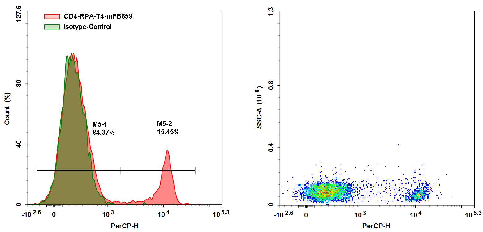
Flow cytometry analysis was performed on whole blood cells stained with mFluor™ Blue 659 anti-human CD4 antibody (Clone: RPA-T4). The fluorescence signal was detected using the NovoCyte 3000 flow cytometer in the PerCP channel.