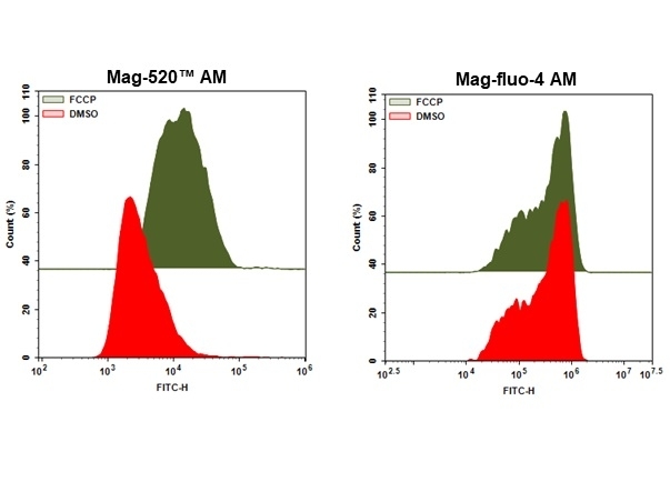
Mag-520™ AM
Intracellular magnesium is important for mediating enzymatic reactions, DNA synthesis, hormone secretion, and muscle contraction. A vast majority of the existing magnesium ion indicators are based on tricarboxylate APTRA chelator derived from the popular tetracarboxylate BAPTA calcium ion chelator. They include mag-fura-2, mag-indo-1, mag-fluo-4 and mag-rhod-3. However, all of them have higher affinity for calcium than magnesium although they were designed for detecting magnesium ion. Mag-520™ is the first commercial magnesium indicator that has higher affinity for magnesium than calcium. Its significantly improved selectivity can be used for magnesium signaling applications with fluorescence microscopy or flow cytometry. Mag-520™ AM is cell-permeable with almost identical spectra to those of FITC, making it a convenient fluorescent probe used with FITC filter set that is ubiquitously equipped with almost all fluorescence instruments.
Example protocol
AT A GLANCE
Protocol summary
- Grow cells as desired
- Prepare and add Mag-520™ AM working solution to samples
- Incubate samples at 37 °C for 15 to 45 minutes
- Monitor the fluorescence intensity using flow cytometer with 530/30 nm filter (FITC channel) or using fluorescence microscopy with FITC filter set
PREPARATION OF STOCK SOLUTIONS
Unless otherwise noted, all unused stock solutions should be divided into single-use aliquots and stored at -20 °C after preparation. Avoid repeated freeze-thaw cycles.
Note Mag-520™ AM working solution should be used promptly.
Mag-520™ AM stock solution
Add appropriate amount of DMSO into Mag-520™ AM vial to make 2-5 mM Mag-520™ AM stock solution.Note Mag-520™ AM working solution should be used promptly.
PREPARATION OF WORKING SOLUTION
Mag-520™ AM working solution
Prepare 5-50 µM Mag-520™ AM working solution in buffer of your choice.Note Mag-520™ AM working solution should be used promptly.
Note The concentration of the Mag-520™ AM should be optimized for different cell types and conditions.
SAMPLE EXPERIMENTAL PROTOCOL
The following protocol can be used as a guideline and should be optimized according to the needs.
- Grow cells as desired.
- Remove the treatment and wash the cells with buffer of your choice such as DPBS.
- Add Mag-520™ AM working solution and incubate the samples for 15-45 minutes at 37 °C incubator.
Note Optimal time for incubation needs to be determined carefully. - Remove the working solution and wash cells with buffer of your choice.
- Resuspend cells in buffer of your choice.
- Add stimulant to stimulate the cells and monitor the fluorescence intensity with flow cytometer using 530/30 nm filter (FITC channel) or fluorescence microscope with FITC filter set.
Calculators
Common stock solution preparation
Table 1. Volume of DMSO needed to reconstitute specific mass of Mag-520™ AM to given concentration. Note that volume is only for preparing stock solution. Refer to sample experimental protocol for appropriate experimental/physiological buffers.
| 0.1 mg | 0.5 mg | 1 mg | 5 mg | 10 mg | |
| 1 mM | 172.574 µL | 862.872 µL | 1.726 mL | 8.629 mL | 17.257 mL |
| 5 mM | 34.515 µL | 172.574 µL | 345.149 µL | 1.726 mL | 3.451 mL |
| 10 mM | 17.257 µL | 86.287 µL | 172.574 µL | 862.872 µL | 1.726 mL |
Molarity calculator
Enter any two values (mass, volume, concentration) to calculate the third.
| Mass (Calculate) | Molecular weight | Volume (Calculate) | Concentration (Calculate) | Moles | ||||
| / | = | x | = |
Spectrum
Citations
View all 15 citations: Citation Explorer
A highly selective fluorescent probe for the intracellular measurement of magnesium ion
Authors: Patel, Deven and Peng, Ruogu and Guo, Haitao and Lin, Raechel and Zhao, Qin and Liao, Jinfang and Diwu, Zhenjun
Journal: Analytical Biochemistry (2020): 113910
Authors: Patel, Deven and Peng, Ruogu and Guo, Haitao and Lin, Raechel and Zhao, Qin and Liao, Jinfang and Diwu, Zhenjun
Journal: Analytical Biochemistry (2020): 113910
A Highly Selective Green Fluorescent Magnesium Indicator for Intracellular Magnesium Ion Analysis
Authors: Patel, Deven and Zhao, Qin and Guo, Haitao and Peng, Ruogu and Liu, Jixiang and Liao, Jinfang and Diwu, Zhenjun
Journal: Biophysical Journal (2020): 273a
Authors: Patel, Deven and Zhao, Qin and Guo, Haitao and Peng, Ruogu and Liu, Jixiang and Liao, Jinfang and Diwu, Zhenjun
Journal: Biophysical Journal (2020): 273a
Fluorescence lifetime imaging of intracellular magnesium content in live cells
Authors: Sargenti, A., C and eo, A., Farruggia, G., D'Andrea, C., Cappadone, C., Malucelli, E., Valentini, G., Taroni, P., Iotti, S.
Journal: Analyst (2019): 1876-1880
Authors: Sargenti, A., C and eo, A., Farruggia, G., D'Andrea, C., Cappadone, C., Malucelli, E., Valentini, G., Taroni, P., Iotti, S.
Journal: Analyst (2019): 1876-1880
Synthesis of a highly Mg(2+)-selective fluorescent probe and its application to quantifying and imaging total intracellular magnesium
Authors: Sargenti, A., Farruggia, G., Zaccheroni, N., Marraccini, C., Sgarzi, M., Cappadone, C., Malucelli, E., Procopio, A., Prodi, L., Lombardo, M., Iotti, S.
Journal: Nat Protoc (2017): 461-471
Authors: Sargenti, A., Farruggia, G., Zaccheroni, N., Marraccini, C., Sgarzi, M., Cappadone, C., Malucelli, E., Procopio, A., Prodi, L., Lombardo, M., Iotti, S.
Journal: Nat Protoc (2017): 461-471
A novel fluorescent chemosensor allows the assessment of intracellular total magnesium in small samples
Authors: Sargenti, A., Farruggia, G., Malucelli, E., Cappadone, C., Merolle, L., Marraccini, C., Andreani, G., Prodi, L., Zaccheroni, N., Sgarzi, M., Trombini, C., Lombardo, M., Iotti, S.
Journal: Analyst (2014): 1201-7
Authors: Sargenti, A., Farruggia, G., Malucelli, E., Cappadone, C., Merolle, L., Marraccini, C., Andreani, G., Prodi, L., Zaccheroni, N., Sgarzi, M., Trombini, C., Lombardo, M., Iotti, S.
Journal: Analyst (2014): 1201-7
Page updated on April 15, 2025


