Indo-1 AM
Product key features
Indo-1 AM is a UV light-excitable, cell-permeable ratiometric calcium indicator used for measuring intracellular calcium-ion concentrations.
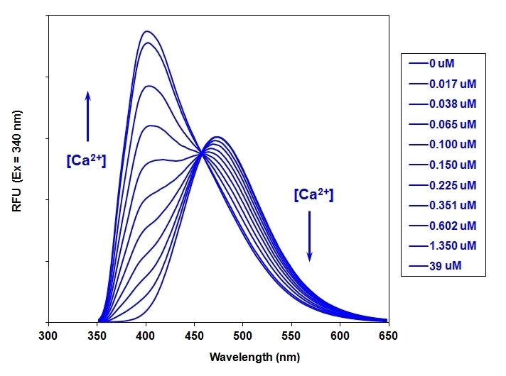
- Ratiometric detection- Distinct shift in emission from 480 nm to 400 nm upon binding to calcium, enabling sensitive and quantitative measurements.
- Optimized for flow cytometry: Requires only a single UV excitation source, such as 351–364 nm spectral lines, simplifying experimental setups.
- Versatile applications: Compatible with various techniques, including fluorescence microscopy, spectroscopy, microplate readers, and flow cytometry.
- Reliable results: Minimal background and superior sensitivity.
Product description
Indo-1 AM is based on the principle of a distinct shift in emission upon binding to Ca²⁺, transitioning from 480 nm (in the absence of Ca²⁺) to 400 nm (when bound to Ca²⁺). Indo-1 AM's high calcium-binding affinity (Kd ~230 nM) makes it highly sensitive to transient calcium ion changes within the cell. Indo-1 AM enables quantitative detection of calcium flux dynamics through real-time emission ratio measurements. The excitation and emission characteristics of Indo-1 AM makes it ideal for multiple applications like fluorescence microscopy, spectroscopy, microplate readers and flow cytometry in particular, as it only requires a single UV excitation source, such as the 351–364 nm lines from an argon-ion laser channel. It delivers superior performance with minimal background, ensuring reliable and reproducible results for complex cellular assays.
Example protocol
PREPARATION OF STOCK SOLUTIONS
Unless otherwise noted, all unused stock solutions should be divided into single-use aliquots and stored at -20 °C after preparation. Avoid repeated freeze-thaw cycles
Prepare a 2 to 5 mM stock solution of Indo-1 AM in high-quality, anhydrous DMSO.
PREPARATION OF WORKING SOLUTION
On the day of the experiment, either dissolve Indo-1 AM in DMSO or thaw an aliquot of the indicator stock solution to room temperature.
Prepare a 2 to 20 µM Indo-1 AM working solution in a buffer of your choice (e.g., Hanks and Hepes buffer) with 0.04% Pluronic® F-127. For most cell lines, Indo-1 AM at a final concentration of 4-5 μM is recommended. The exact concentration of indicators required for cell loading must be determined empirically.
Note: The nonionic detergent Pluronic® F-127 is sometimes used to increase the aqueous solubility of Indo-1 AM. A variety of Pluronic® F-127 solutions can be purchased from AAT Bioquest.
Note: If your cells contain organic anion-transporters, probenecid (1-2 mM) may be added to the dye working solution (final in well concentration will be 0.5-1 mM) to reduce leakage of the de-esterified indicators. A variety of ReadiUse™ Probenecid products, including water-soluble, sodium salt, and stabilized solutions, can be purchased from AAT Bioquest.
SAMPLE EXPERIMENTAL PROTOCOL
Following is our recommended protocol for loading AM esters into live cells. This protocol only provides a guideline and should be modified according to your specific needs.
- Prepare cells in growth medium overnight.
On the next day, add 1X Indo-1 AM working solution to your cell plate.
Note: If your compound(s) interfere with the serum, replace the growth medium with fresh HHBS buffer before dye-loading.
Incubate the dye-loaded plate in a cell incubator at 37 °C for 30 to 60 minutes.
Note: Incubating the dye for longer than 1 hour can improve signal intensities in certain cell lines.
- Replace the dye working solution with HHBS or buffer of your choice (containing an anion transporter inhibitor, such as 1 mM probenecid, if applicable) to remove any excess probes.
- Add the stimulant as desired and simultaneously measure fluorescence using either a fluorescence microscope equipped with an Indo-1 filter set or a fluorescence plate reader containing a programmable liquid handling system such as a FlexStation, at Ex/Em1 = 340/400 nm no cutoff and Ex/Em2 = 340/475 cutoff 455 nm.
Calculators
Common stock solution preparation
0.1 mg | 0.5 mg | 1 mg | 5 mg | 10 mg | |
1 mM | 99.019 µL | 495.094 µL | 990.187 µL | 4.951 mL | 9.902 mL |
5 mM | 19.804 µL | 99.019 µL | 198.037 µL | 990.187 µL | 1.98 mL |
10 mM | 9.902 µL | 49.509 µL | 99.019 µL | 495.094 µL | 990.187 µL |
Molarity calculator
Mass (Calculate) | Molecular weight | Volume (Calculate) | Concentration (Calculate) | Moles | ||||
/ | = | x | = |
Spectrum
Citations
Authors: Li, Jiuru and Wiesinger, Alexandra and Fokkert, Lianne and Bakker, Priscilla and de Vries, Dylan K and Tijsen, Anke J and Pinto, Yigal M and Verkerk, Arie O and Christoffels, Vincent M and Boink, Gerard JJ and others,
Journal: Cell Stem Cell (2024)
Authors: Birkner, Antje
Journal: (2019)
Authors: Zhao, Tao and Guo, Dongqing and Gu, Yuchun and Ling, Yang
Journal: Molecular Medicine Reports (2017): 2259--2263
Authors: Guo, Dongqing and Gu, Junzhong and Jiang, Hui and Ahmed, Asif and Zhang, Zhiren and Gu, Yuchun
Journal: Journal of molecular and cellular cardiology (2016): 179--187
Authors: Guo, Dong-Qing and Zhang, Hao and Tan, Sheng-Jiang and Gu, Yu-Chun
Journal: PloS one (2014): e113649
References
Authors: Nelemans A., undefined
Journal: Methods Mol Biol (2006): 47
Authors: Bailey S, Macardle PJ.
Journal: J Immunol Methods (2006): 220
Authors: Eerbeek O, Mik EG, Zuurbier CJ, van 't Loo M, Donkersloot C, Ince C.
Journal: J Appl Physiol (2004): 2042
Authors: Sakurai K, Norota I, Tanaka H, Kubota I, Tomoike H, Endo M.
Journal: Life Sci (2002): 1173
Authors: Endoh M., undefined
Journal: Nippon Yakurigaku Zasshi (2000): 361