logo
AAT Bioquest

CytoTell™ Blue

Flow cytometry combined with fluorescence staining is a powerful tool to analyze heterogeneous cell populations. Among all the existing fluorescent dyes CFSE is the preferred cell proliferation indicator that is widely used for live cell analysis. However, it is impossible to use CFSE and its fluorescein analogs for GFP-transfected cells or for the applications where a FITC-labeled antibody is used since CFSE and its fluorescein analogs have the excitation and emission spectra almost identical to GFP or FITC. CytoTell™ dyes are well excited at major laser lines such as 405 nm, 488 nm or 633 nm with multicolor emissions. CytoTell™ dyes have minimal cytotoxicity, and are used for the multicolor applications with either GFP cell lines or FITC-labeled antibodies since they have either excitation or emission spectra distinct from fluorescein. CytoTell™ Blue is a blue fluorescent dye that stains cells evenly. As cells divide, the dye is distributed equally between daughter cells that can be measured as successive halving of the fluorescence intensity of the dye. Cells labeled with CytoTell™ Blue may be fixed and permeabilized for analysis of intracellular targets using standard formaldehyde-containing fixatives and saponin-based permeabilization buffers. CytoTell™ Blue has a peak excitation of 405 nm and can be excited by the violet (405 nm) laser line. It has a peak emission of 450 nm and can be detected with a 450/20 band pass filter (equivalent to Pacific Blue®, or BD Horizon® V450), making it compatible with applications that utilize GFP or FITC antibodies for multicolor cell analysis.
Example protocol

AT A GLANCE

Protocol summary

  1. Prepare cells with test compounds
  2. Add 1X dye working solution
  3. Incubate dyes with cells at room temperature or 37 oC for 10 to 30 minutes
  4. Remove the dye working solution
  5. Analyse with flow cytometer with appropriate filter set

Important notes
Bring all the kit components at room temperature before starting the experiment. Note: The CytoTell™ dyes are lyophilized powders. They should be stable for at least 6 months if store at -20 °C, protecting from light, and avoiding freeze/thaw cycles.

Product Number

Indicator

Size

Ex/Em (nm)

Excitation Source

22240

CytoTell™ UltraGreen

500 tests

492/519

488 nm (Blue Laser)

22241

CytoTell™ UltraGreen

1000 tests

492/519

488 nm (Blue Laser)

22248

CytoTell™ Violet 500

500 tests

415/499

405 nm (Violet Laser)

22251

CytoTell™ Blue

500 tests

403/454

405 nm (Violet Laser)

22252

CytoTell™ Blue

1000 tests

403/454

405 nm (Violet Laser)

22253

CytoTell™ Green

500 tests

511/525

488 nm (Blue Laser)

22254

CytoTell™ Green

1000 tests

511/525

488 nm (Blue Laser)

22255

CytoTell™ Red 650

500 tests

628/643

633 nm (Red Laser)

22256

CytoTell™ Red 650

1000 tests

628/643

633 nm (Red Laser)

22257

CytoTell™ Orange

500 tests

542 /556

488 nm (Blue Laser)
531 nm (Green Laser)

22258

CytoTell™ Orange

1000 tests

542 /556

488 nm (Blue Laser)
531 nm (Green Laser)

22261

CytoTell™ Red 590

500 tests

560 /574

488 nm (Blue Laser)
531 nm (Green Laser)

22262

CytoTell™ Red 590

1000 tests

560 /574

488 nm (Blue Laser)
531 nm (Green Laser)

PREPARATION OF STOCK SOLUTION

Unless otherwise noted, all unused stock solutions should be divided into single-use aliquots and stored at -20 °C after preparation. Avoid repeated freeze-thaw cycles.

CytoTell™ dye stock solution (500X):
Add 500 µL DMSO into the dye powder vial, mix it well by vortexing to have a stock solution (500X). Note: The stock solution should be used promptly; any remaining solution should be aliquoted and frozen at < - 20 oC. Avoid repeated freeze-thaw cycles, and protect from light.

PREPARATION OF WORKING SOLUTION

CytoTellTM dye working solution (1X):
Dilute the 500X DMSO stock solution at 1 to 500 in Hanks and 20 mM Hepes buffer (HHBS) or the buffer of your choice, pH 7 (such as 1 µL of 500X DMSO stock solution to 500 µL buffer) right before use. Mix them well by vortexing. Note: The final concentration of the dye working solution should be empirically determined for different cell types and/or experimental conditions. It is recommended to test at the concentrations that are at least over ten fold range. Such as CytoTell™ Red might use much less amount in some cell types than the recommend concentrations.

SAMPLE EXPERIMENTAL PROTOCOL

  1. Treat cells with test compounds for a desired period of time.

  2. Centrifuge the cells to get 1-5 × 10⁵ cells per tube.

  3. Resuspend cells in 500 µL of the CytoTell™ dye working solution. Optional: One can add the 500X DMSO stock solution into the cells directly without medium removing (such as, add 1 µL500X DMSO stock solution into 500 µL cells)

  4. Incubate cells with a dye solution at room temperature or 37 °C for 10 to 30 minutes, protected from light.

  5. Remove the dye working solution from the cells, wash the cells with HHBS or buffer of your choice. Resuspend cells in 500 µL of pre-warmed HHBS or medium to get 1-5 × 10⁵ cells per tube.

  6. Monitor the fluorescence change at respected Ex/Em (see Table 1) with a flow cytometer or a fluorescence microscope.

Calculators

Common stock solution preparation

Table 1. Volume of DMSO needed to reconstitute specific mass of CytoTell™ Blue to given concentration. Note that volume is only for preparing stock solution. Refer to sample experimental protocol for appropriate experimental/physiological buffers.

0.1 mg0.5 mg1 mg5 mg10 mg
1 mM263.359 µL1.317 mL2.634 mL13.168 mL26.336 mL
5 mM52.672 µL263.359 µL526.718 µL2.634 mL5.267 mL
10 mM26.336 µL131.679 µL263.359 µL1.317 mL2.634 mL

Molarity calculator

Enter any two values (mass, volume, concentration) to calculate the third.

Mass (Calculate)Molecular weightVolume (Calculate)Concentration (Calculate)Moles
/=x=

Spectrum

Product family

NameExcitation (nm)Emission (nm)
CytoTell™ Green510525
CytoTell™ Orange541560

Citations

View all 17 citations: Citation Explorer
The CH1 domain influences the expression and antigen sensing of the HIV-specific CH31 IgM-BCR and IgG-BCR
Authors: Ortiz, Yaneth and Anasti, Kara and Pane, Advaiti K and Cronin, Kenneth and Alam, S Munir and Reth, Michael
Journal: Proceedings of the National Academy of Sciences (2024): e2404728121
Vaccine plus microbicide effective in preventing vaginal SIV transmission in macaques
Authors: Rahman, MA and Bissa, M and Silva de Castro, I and Helmold Hait, S and Stamos, JD and Bhuyan, F and Hunegnaw, R and Sarkis, S and Gutowska, A and Doster, MN and others,
Journal: Nature Microbiology (2023): 1--14
The impact of CCR8+ regulatory T cells on cytotoxic T cell function in human lung cancer
Authors: Haruna, Miya and Ueyama, Azumi and Yamamoto, Yoko and Hirata, Michinari and Goto, Kumiko and Yoshida, Hiroshi and Higuchi, Naoko and Yoshida, Tetsuya and Kidani, Yujiro and Nakamura, Yamami and others,
Journal: Scientific Reports (2022): 1--12
Omega-3 docosahexaenoic acid (DHA) impedes silica-induced macrophage corpse accumulation by attenuating cell death and potentiating efferocytosis
Authors: Rajasinghe, Lichchavi D and Chauhan, Preeti S and Wierenga, Kathryn A and Evered, Augustus O and Harris, Shamya N and Bates, Melissa A and Gavrilin, Mikhail A and Pestka, James J
Journal: Frontiers in immunology (2020): 2179
PD-1+ Tim3+ tumor-infiltrating CD8 T cells sustain the potential for IFN-$\gamma$ production, but lose cytotoxic activity in ovarian cancer
Authors: Sawada, Masaaki and Goto, Kumiko and Morimoto-Okazawa, Akiko and Haruna, Miya and Yamamoto, Kei and Yamamoto, Yoko and Nakagawa, Satoshi and Hiramatsu, Kosuke and Matsuzaki, Shinya and Kobayashi, Eiji and others,
Journal: International Immunology (2020): 397--405

References

View all 50 references: Citation Explorer
Phenotyping of circulating CD8(+) T cell subsets in human cutaneous leishmaniasis
Authors: Khamesipour A, Nateghi Rostami M, Tasbihi M, Miramin Mohammadi A, Shahrestani T, Sarrafnejad A, Sohrabi Y, Esk and ari SE, Keshavarz Valian H.
Journal: Microbes Infect (2012): 702
Evaluation of multitype mathematical models for CFSE-labeling experiment data
Authors: Miao H, Jin X, Perelson AS, Wu H.
Journal: Bull Math Biol (2012): 300
Repetitive transplantation of different cell types sequentially improves heart function after infarction
Authors: Alex, undefined and er S, Sasse A, Konschalla S, Kroh A, Merx MW, Weber C, Liehn EA.
Journal: J Cell Mol Med (2012): 1640
Identification and isolation of slow-dividing cells in human glioblastoma using carboxy fluorescein succinimidyl ester (CFSE)
Authors: Deleyrolle LP, Rohaus MR, Fortin JM, Reynolds BA, Azari H.
Journal: J Vis Exp. (2012)
New and improved methods for measuring lymphocyte proliferation in vitro and in vivo using CFSE-like fluorescent dyes
Authors: Quah BJ, Parish CR.
Journal: J Immunol Methods (2012): 1
Page updated on November 20, 2024

Ordering information

Price
Unit size
500 Tests
2x500 Tests
Catalog Number
2225122252
Quantity
Add to cart

Additional ordering information

Telephone1-800-990-8053
Fax1-800-609-2943
Emailsales@aatbio.com
InternationalSee distributors
Bulk requestInquire
Custom sizeInquire
Technical SupportContact us
Purchase orderSend to sales@aatbio.com
ShippingStandard overnight for United States, inquire for international
Request quotation

Physical properties

Molecular weight

379.71

Solvent

DMSO

Spectral properties

Excitation (nm)

410

Emission (nm)

445

Storage, safety and handling

H-phraseH303, H313, H333
Hazard symbolXN
Intended useResearch Use Only (RUO)
R-phraseR20, R21, R22

Storage

Freeze (< -15 °C); Minimize light exposure
UNSPSC12352200

Platform

Flow cytometer

Excitation405 nm laser
Emission450, 40 nm filter
Instrument specification(s)Pacific Blue channel
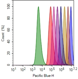
Cell proliferation assay with CytoTell&trade;Blue. Jurkat cells are stained with&nbsp; CytoTell&trade;&nbsp;Blue on Day0, and serially passed at 1:1 ratio for 8 days. Fluorescence intensity of each generation was measured with ACEA&nbsp; NovoCyte 3000 flow cytometer Pacific Blue channel.