Cell Meter™ Generic Fluorimetric Caspase Activity Assay Kit *Green Fluorescence Optimized for Flow Cytometry*
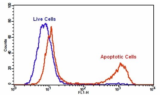
Our Cell Meter™ assay kits are a set of tools for monitoring cell viability. There are a variety of parameters that can be used for monitoring cell viability. This particular kit is designed to monitor cell apoptosis through measuring generic caspases (caspase-1, -3, -4, -5, -6, -7, -8 and -9) activation in living cells. Caspases activation is widely accepted as a reliable indicator for cell apoptosis. Most caspases have substrate selectivity for the peptide sequence Val-Ala-Asp (VAD). This kit uses TF2-VAD-FMK as a fluorogenic indicator for most caspase activity. TF2-VAD-FMK is cell permeable, nontoxic, and irreversibly binds to activated casepase-1, -3, -4, -5, -6, -7, -8 and -9 in apoptotic cells. Once bound to caspases, the green fluorescent reagent is retained within the cell. The binding event prevents the caspases from further catalysis but will not stop apoptosis from proceeding. The reagent will start to react with active caspase enzymes within 15 minutes of addition to the media. The kit provides all the essential components with an optimized assay protocol. It is used for the quantification of most activated caspases activities in apoptotic cells, or for screening caspases inhibitors. The green label allows for direct detection of activated caspases in apoptotic cells by flow cytometry.
Example protocol
AT A GLANCE
Protocol summary
- Prepare cells with test compounds at a density of 5 × 10⁵ to 1 × 10⁶ cells/mL
- Add 1 µL of 500X TF2-VAD-FMK into 0.5 mL of cell solution
- Incubate the cells in a 37°C, 5% CO2 incubator for 1 - 4 hours
- Pellet the cells and resuspend the cells in 0.5 mL of assay buffer or growth medium
- Analyze cells using flow cytometer with 530/30 nm filter (FITC channel)
Important notes
Thaw all the kit components at room temperature before starting the experiment.
SAMPLE EXPERIMENTAL PROTOCOL
- For each sample, prepare cells in 0.5 mL warm medium or buffer of your choice at a density of 5 × 10⁵ to 1 × 10⁶ cells/mL. Note: Each cell line should be evaluated on an individual basis to determine the optimal cell density for apoptosis induction.
- Treat cells with test compounds for a desired period of time to induce apoptosis, and create positive and negative controls.
- Add 1 µL of 500X TF2-VAD-FMK (Component A) into the treated cells.
- Incubate the cells in a 37°C, 5% CO2 incubator for 1 - 4 hours. Note: For adherent cells, gently lift the cells with 0.5 mM EDTA to keep the cells intact, and wash the cells once with serum-containing media prior to incubation with TF2-VAD-FMK. The appropriate incubation time depends on the individual cell type and cell concentration used. Optimize the incubation time for each experiment.
- Wash and spin the cells twice. Resuspend the cells in 0.5 mL of Assay Buffer (Component B) or growth medium. Note: TF2-VAD-FMK is fluorescent; therefore it is important to wash out any unbound reagent to remove the background.
- If desired, label the cells with a DNA stain (such as propidium iodide or 7-AAD for dead cells).
- If desired, fix cells.
- Monitor the fluorescence intensity using a flow cytometer wih 530/30 nm filter (FITC channel). Gate on the cells of interest, excluding debris.
Spectrum
Open in Advanced Spectrum Viewer
Product family
Name | Excitation (nm) | Emission (nm) | Extinction coefficient (cm -1 M -1) | Quantum yield | Correction Factor (280 nm) |
Cell Meter™ Generic Fluorimetric Caspase Binding Assay Kit *Red Fluorescence Optimized for Flow Cytometry* | 649 | 663 | 250000 | 0.271 | 0.027 |
Citations
View all 16 citations: Citation Explorer
Extract of Juniperus indica Bertol synergizes with cisplatin to inhibit oral cancer cell growth via repression of cell cycle progression and activation of the caspase cascade
Authors: Huang, Xiao-Fan and Chang, Kai-Fu and Lee, Shan-Chih and Li, Chia-Yu and Liao, Hung-Hsiu and Hsieh, Ming-Chang and Tsai, Nu-Man
Journal: Molecules (2020): 2746
Authors: Huang, Xiao-Fan and Chang, Kai-Fu and Lee, Shan-Chih and Li, Chia-Yu and Liao, Hung-Hsiu and Hsieh, Ming-Chang and Tsai, Nu-Man
Journal: Molecules (2020): 2746
Promotion of osteointegration under diabetic conditions by tantalum coating-based surface modification on 3-dimensional printed porous titanium implants
Authors: Wang, Lin and Hu, Xiaofan and Ma, Xiangyu and Ma, Zhensheng and Zhang, Yang and Lu, Yizhao and Li, Xiang and Lei, Wei and Feng, Yafei
Journal: Colloids and Surfaces B: Biointerfaces (2016): 440--452
Authors: Wang, Lin and Hu, Xiaofan and Ma, Xiangyu and Ma, Zhensheng and Zhang, Yang and Lu, Yizhao and Li, Xiang and Lei, Wei and Feng, Yafei
Journal: Colloids and Surfaces B: Biointerfaces (2016): 440--452
microRNA-186 inhibits cell proliferation and induces apoptosis in human esophageal squamous cell carcinoma by targeting SKP2
Authors: He, Wei and Feng, Jianfang and Zhang, Yan and Wang, Yuanyuan and Zang, Wenqiao and Zhao, Guoqiang
Journal: Laboratory Investigation (2016): 317--324
Authors: He, Wei and Feng, Jianfang and Zhang, Yan and Wang, Yuanyuan and Zang, Wenqiao and Zhao, Guoqiang
Journal: Laboratory Investigation (2016): 317--324
Hypoxia regulates TRAIL sensitivity of colorectal cancer cells through mitochondrial autophagy.
Authors: Knoll, Gertrud and Bittner, Sebastian and Kurz, Maria and Jantsch, Jonathan and Ehrenschwender, Martin
Journal: Oncotarget (2016)
Authors: Knoll, Gertrud and Bittner, Sebastian and Kurz, Maria and Jantsch, Jonathan and Ehrenschwender, Martin
Journal: Oncotarget (2016)
Role of delta-like ligand-4 in chemoresistance against docetaxel in MCF-7 cells
Authors: Wang, Q and Shi, Y and Butler, HJ and Xue, J and Wang, G and Duan, P and Zheng, H
Journal: Human & Experimental Toxicology (2016): 0960327116650006
Authors: Wang, Q and Shi, Y and Butler, HJ and Xue, J and Wang, G and Duan, P and Zheng, H
Journal: Human & Experimental Toxicology (2016): 0960327116650006
References
View all 46 references: Citation Explorer
Evidence for programmed cell death and activation of specific caspase-like enzymes in the tomato fruit heat stress response
Authors: Qu GQ, Liu X, Zhang YL, Yao D, Ma QM, Yang MY, Zhu WH, Yu S, Luo YB.
Journal: Planta (2009): 1269
Authors: Qu GQ, Liu X, Zhang YL, Yao D, Ma QM, Yang MY, Zhu WH, Yu S, Luo YB.
Journal: Planta (2009): 1269
Gamma-linolenic acid induces apoptosis and lipid peroxidation in human chronic myelogenous leukemia K562 cells
Authors: Ge H, Kong X, Shi L, Hou L, Liu Z, Li P.
Journal: Cell Biol Int (2009): 402
Authors: Ge H, Kong X, Shi L, Hou L, Liu Z, Li P.
Journal: Cell Biol Int (2009): 402
Apoptosis induced by enniatins H and MK1688 isolated from Fusarium oxysporum FB1501
Authors: Hyun U, Lee DH, Lee C, Shin CG.
Journal: Toxicon (2009): 723
Authors: Hyun U, Lee DH, Lee C, Shin CG.
Journal: Toxicon (2009): 723
Suppression of autophagy enhances the cytotoxicity of the DNA-damaging aromatic amine p-anilinoaniline
Authors: Elliott A, Reiners JJ, Jr.
Journal: Toxicol Appl Pharmacol (2008): 169
Authors: Elliott A, Reiners JJ, Jr.
Journal: Toxicol Appl Pharmacol (2008): 169
Beta-sitosterol sensitizes MDA-MB-231 cells to TRAIL-induced apoptosis
Authors: Park C, Moon DO, Ryu CH, Choi B, Lee W, Kim GY, Choi Y.
Journal: Acta Pharmacol Sin (2008): 341
Authors: Park C, Moon DO, Ryu CH, Choi B, Lee W, Kim GY, Choi Y.
Journal: Acta Pharmacol Sin (2008): 341
Page updated on December 17, 2024