logo
AAT Bioquest

Cell Meter™ Fluorimetric Mitochondrial Superoxide Activity Assay Kit*Optimized for Microplate Reader*

Mitochondria are major producers of cellular superoxide. The production of low to moderate levels of superoxide is critical for the proper regulation of many essential cellular processes including gene expression, signal transduction, and muscle adaptation to endurance exercise training. Uncontrolled mitochondrial superoxide production can trigger cellular oxidative damage that contributes to the pathogenesis of a wide variety of disorders including cancer, cardiovascular diseases, neurodegenerative diseases and aging. The detection of intracellular mitochondrial superoxide is of central importance to understanding proper cellular redox regulation and the impact of its dysregulation on various pathologies. Cell Meter™ Fluorimetric Mitochondrial Superoxide Activity Assay Kit uses our unique Superoxide Indicator to quantify superoxide level in live cells. MitoROS™ 580 is live-cell permeant and can rapidly and selectively target superoxide in mitochondria. It generates red fluorescence when it reacts with superoxide. The Cell Meter™ Fluorimetric Intracellular Superoxide Detection Kit provides a sensitive, one-step fluorimetric assay to detect mitochondrial superoxide in live cells with one hour incubation. This kit can be used for fluorescence microplate readers and fluorescence microscopy applications.
Example protocol

AT A GLANCE

Protocol summary

  1. Prepare cells in growth medium
  2. Treat the cells with test compounds to induce superoxide
  3. Add MitoROS™ 580 working solution
  4. Stain the cells at 37°C for 30 - 60 minutes
  5. Monitor the fluorescence increase (bottom read mode) at Ex/Em= 540/590 nm (Cutoff = 570 nm) or fluorescence microscope with TRITC filter set

Important notes
Thaw all the components at room temperature before starting the experiment.

PREPARATION OF STOCK SOLUTION

Unless otherwise noted, all unused stock solutions should be divided into single-use aliquots and stored at -20 °C after preparation. Avoid repeated freeze-thaw cycles.

1. MitoROS™ 580 stock solution (500X):
Add 50 µL of DMSO (Component C) into the vial of MitoROS™ 580 (Component A) and mix well to make 500X MitoROS™ 580 stock solution. Protect from light. Note: 25 µL of 500X MitoROS™ 580 stock solution is enough for 1 plate. For storage, seal tubes tightly.

PREPARATION OF WORKING SOLUTION

Add 25 μL of 500X MitoROS™ 580 stock solution into 10 mL of Assay Buffer (Component B) and mix well to make MitoROS™ 580 working solution. Note: This MitoROS™ 580 working solution is stable for at least 2 hours at room temperature.

For guidelines on cell sample preparation, please visit
https://www.aatbio.com/resources/guides/cell-sample-preparation.html

SAMPLE EXPERIMENTAL PROTOCOL

  1. Treat cells with 10 µL of 10X test compounds (96-well plate) or 5 µL of 5X test compounds (384-well plate) in your desired buffer (such as PBS or HHBS). For control wells (untreated cells), add the corresponding amount of compound buffer.

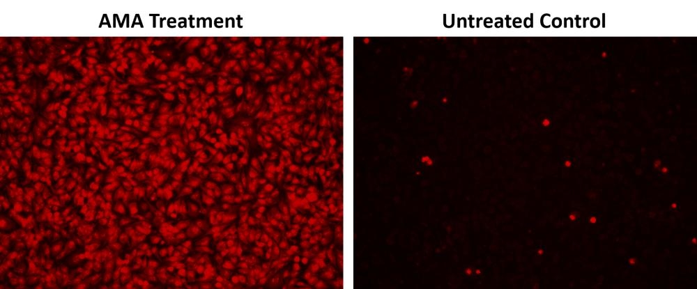
  2. To induce superoxide, incubate the cell plate at 37°C for a desired period of time, protect from light. Note: We treated HeLa cells with 50 µM Antimycin A (AMA) at 37°C for 30 minutes to induce superoxide. See Figure 1 for details.

  3. Add 100 µL/well (96-well plate) or 25 µL/well (384-well plate) of MitoROS™ 580 working solution into the cell plate.

  4. Incubate the cells at 37°C for 30 to 60 minutes.

  5. Monitor the fluorescence increase with a fluorescence microplate reader (bottom read mode) at  Ex/Em = 540/590 nm (Cutoff = 570 m) or observe cells using a fluorescence microscope with TRITC filter.

Citations

View all 5 citations: Citation Explorer
Mitochondrial genome transfer drives metabolic reprogramming in adjacent colonic epithelial cells promoting TGF$\beta$1-mediated tumor progression
Authors: Guan, Bingjie and Liu, Youdong and Xie, Bowen and Zhao, Senlin and Yalikun, Abudushalamu and Chen, Weiwei and Zhou, Menghua and Gu, Qi and Yan, Dongwang
Journal: Nature Communications (2024): 1--18
GRP75-faciliated Mitochondria-associated ER Membrane (MAM) Integrity controls Cisplatin-resistance in Ovarian Cancer Patients
Authors: Li, Jing and Qi, Fangzheng and Su, Huishan and Zhang, Chuanshan and Zhang, Qing and Chen, Ying and Chen, Ping and Su, Linjia and Chen, Yanan and Yang, Yuqi and others,
Journal: International journal of biological sciences (2022): 2914
Effect of antioxidants on the H2O2-induced premature senescence of human fibroblasts
Authors: Pie{\'n}kowska, Natalia and Bartosz, Grzegorz and Pichla, Monika and Grzesik-Pietrasiewicz, Michalina and Gruchala, Martyna and Sadowska-Bartosz, Izabela
Journal: Aging (Albany NY) (2020): 1910
Nitroxide Radical-Containing Redox Nanoparticles Protect Neuroblastoma SH-SY5Y Cells against 6-Hydroxydopamine Toxicity
Authors: Pichla, Monika and Pulaski, {\L}ukasz and Kania, Katarzyna Dominika and Stefaniuk, Ireneusz and Cieniek, Bogumi{\l} and Pie{\'n}kowska, Natalia and Bartosz, Grzegorz and Sadowska-Bartosz, Izabela
Journal: Oxidative Medicine and Cellular Longevity (2020)

References

View all 60 references: Citation Explorer
Concentration-dependent effect of sodium hypochlorite on stem cells of apical papilla survival and differentiation
Authors: Martin DE, De Almeida JF, Henry MA, Khaing ZZ, Schmidt CE, Teixeira FB, Diogenes A.
Journal: J Endod (2014): 51
Effect of hypochlorite oxidation on cholinesterase-inhibition assay of acetonitrile extracts from fruits and vegetables for monitoring traces of organophosphate pesticides
Authors: Kitamura K, Maruyama K, Hamano S, Kishi T, Kawakami T, Takahashi Y, Onodera S.
Journal: J Toxicol Sci (2014): 71
Green synthesis of carbon dots with down- and up-conversion fluorescent properties for sensitive detection of hypochlorite with a dual-readout assay
Authors: Yin B, Deng J, Peng X, Long Q, Zhao J, Lu Q, Chen Q, Li H, Tang H, Zhang Y, Yao S.
Journal: Analyst (2013): 6551
Comparative antimicrobial activities of aerosolized sodium hypochlorite, chlorine dioxide, and electrochemically activated solutions evaluated using a novel standardized assay
Authors: Thorn RM, Robinson GM, Reynolds DM.
Journal: Antimicrob Agents Chemother (2013): 2216
Analysis of the germination kinetics of individual Bacillus subtilis spores treated with hydrogen peroxide or sodium hypochlorite
Authors: Setlow B, Yu J, Li YQ, Setlow P.
Journal: Lett Appl Microbiol (2013): 259
Page updated on November 21, 2024

Ordering information

Price
Unit size
Catalog Number22971
Quantity
Add to cart

Additional ordering information

Telephone1-800-990-8053
Fax1-800-609-2943
Emailsales@aatbio.com
InternationalSee distributors
Bulk requestInquire
Custom sizeInquire
Technical SupportContact us
Purchase orderSend to sales@aatbio.com
ShippingStandard overnight for United States, inquire for international
Request quotation

Storage, safety and handling

H-phraseH303, H313, H333
Hazard symbolXN
Intended useResearch Use Only (RUO)
R-phraseR20, R21, R22
UNSPSC12352200

Platform

Fluorescence microplate reader

Excitation540 nm
Emission590 nm
Cutoff570 nm
Recommended plateBlack wall, clear bottom
Instrument specification(s)Bottom read mode

Components

Fluorescence images of superoxide measurement in HeLa cells using Cell Meter™ Fluorimetric Intracellular Superoxide Detection Kit (Cat#22971). HeLa cells at 100,000 cells/well/100 µL were seeded overnight in a 96-well black wall/clear bottom plate. AMA Treatment: Cells were treated with 50 µM Antimycin A (AMA) at 37 °C for 30 minutes, then incubated with MitoROS™ 580 for 1 hour. Untreated Control: HeLa cells were incubated with MitoROS™ 580 at 37 °C for 1 hour without AMA treatment. The fluorescence signal was measured using fluorescence microscope with a TRITC filter.
Detection of intracellular superoxide in HeLa cells using Cell Meter™ Fluorimetric Intracellular Superoxide Detection Kit (Cat#22971). HeLa cells at 100,000 cells/well/100 µL were seeded overnight in a 96-well black wall/clear bottom plate. Cells were incubated with 50 µM Pyocyanin (Pyo); 50 µM Antimycin A (AMA) or without treatment (Control) at 37 ºC for 30 minutes. Cells were then incubated with MitoROS™ 580 at 37 ºC for 1 hour. The fluorescence signal were monitored at Ex/Em = 540/590 nm (Cutoff = 570 nm) with bottom read mode using a CLARIOstar microplate reader (BMG Labtech).