logo
AAT Bioquest

Cell Meter™ Fluorimetric Intracellular Total ROS Activity Assay Kit*Green Fluorescence*

Reactive oxygen species (ROS) are natural byproducts of the normal metabolism of oxygen and play important roles in cell signaling. However, during oxidative stress-related states, ROS levels can increase dramatically. The accumulation of ROS results in significant damage to cell structures. The role of oxidative stress in cardiovascular disease, diabetes, osteoporosis, stroke, inflammatory diseases, a number of neurodegenerative diseases and cancer has been well established. The ROS measurement will help to determine how oxidative stress modulates varied intracellular pathways. Cell Meter™ Fluorimetric ROS Assay Kit uses our unique ROS sensor to quantify ROS in live cells. ROS Green is cell-permeable. It generates the green fluorescence when it reacts with ROS. The kit is an optimized "mix and read" assay format that is compatible with HTS liquid handling instruments. The Cell Meter™ Fluorimetric ROS Assay Kit provides a sensitive, one-step fluorimetric assay to detect intracellular ROS in live cells with one hour incubation. The assay can be performed in a convenient 96-well or 384-well microtiter-plate format and easily adapted to automation without a separation step. Its signal can be easily read using either a fluorescence microplate reader or a fluorescence microscope.
Example protocol

AT A GLANCE

Protocol summary

  1. Prepare cells in growth medium
  2. Add Amplite™ ROS Green working solution (100 µL/well for a 96- well plate or 25 µL/well for a 384-well plate) 
  3. Stain the cells at 37°C for 60 minutes
  4. Treat the cells with test compounds to induce ROS
  5. Monitor the fluorescence increase (bottom read mode) at Ex/Em= 490/525 nm (Cutoff = 515 nm) or fluorescence microscope with FITC filter set

Important notes
Thaw all the kit components at room temperature before starting the experiment.

PREPARATION OF STOCK SOLUTION

Unless otherwise noted, all unused stock solutions should be divided into single-use aliquots and stored at -20 °C after preparation. Avoid repeated freeze-thaw cycles.

1. Amplite™ ROS Green stock solution (500X):
Add 40 µL of DMSO (Component C) into the vial of Amplite™ ROS Green (Component A) and mix well to make 500X Amplite™ ROS Green stock solution. Protect from light. Note: 20 µL of 500X Amplite™ ROS Green stock solution is enough for 1 plate. For storage, seal tubes tightly.

PREPARATION OF WORKING SOLUTION

Add 20 µL of 500X Amplite™ ROS Green stock solution into 10 mL of Assay Buffer (Component B) and mix well to make Amplite™ ROS Green working solution. Note: This Amplite™ ROS Green working solution is stable for at least 2 hours at room temperature.

For guidelines on cell sample preparation, please visit
https://www.aatbio.com/resources/guides/cell-sample-preparation.html

SAMPLE EXPERIMENTAL PROTOCOL

  1. Add 100 µL/well (96-well plate) or 25 µL/well (384-well plate) of Amplite™ ROS Green working solution into the cell plate.

  2. Incubate the cells in a 5% CO2, 37°C incubator for one hour.

  3. Treat cells with 20 µL of 11X test compounds (96-well plate) or 10 µL of 6X test compounds (384-well plate) in your desired buffer (such as PBS or HHBS). For control wells (untreated cells), add the corresponding amount of compound buffer.

  4. To induce ROS, incubate the cell plate at room temperature or in a 5% CO2, 37°C incubator for at least 15 minutes or a desired period of time (30 minutes for Hela cells treated with 1 mM H2O2).

  5. Monitor the fluorescence increase with a fluorescence microplate reader (bottom read mode) at Ex/Em = 490/525 nm (Cutoff = 515 nm) or observe cells using a fluorescence microscope with FITC filter set.

Citations

View all 34 citations: Citation Explorer
Palmitic Acid Exerts Anti-Tumorigenic Activities by Modulating Cellular Stress and Lipid Droplet Formation in Endometrial Cancer
Authors: Zhao, Ziyi and Wang, Jiandong and Kong, Weimin and Newton, Meredith A and Burkett, Wesley C and Sun, Wenchuan and Buckingham, Lindsey and O’Donnell, Jillian and Suo, Hongyan and Deng, Boer and others,
Journal: Biomolecules (2024): 601
Linoleic acid exhibits anti-proliferative and anti-invasive activities in endometrial cancer cells and a transgenic model of endometrial cancer
Authors: Qiu, Jianqing and Zhao, Ziyi and Suo, Hongyan and Paraghamian, Sarah E and Hawkins, Gabrielle M and Sun, Wenchuan and Zhang, Xin and Hao, Tianran and Deng, Beor and Shen, Xiaochang and others,
Journal: Cancer Biology \& Therapy (2024): 2325130
Shank3 ameliorates neuronal injury after cerebral ischemia/reperfusion via inhibiting oxidative stress and inflammation
Authors: Zhang, Hongchen and Feng, Yuan and Si, Yanfang and Lu, Chuanhao and Wang, Juan and Wang, Shiquan and Li, Liang and Xie, Wenyu and Yue, Zheming and Yong, Jia and others,
Journal: Redox Biology (2023): 102983
Reduced expression of phosphorylated ataxia-telangiectasia mutated gene is related to poor prognosis and gemcitabine chemoresistance in pancreatic cancer
Authors: Xun, Jingyu and Ohtsuka, Hideo and Hirose, Katsuya and Douchi, Daisuke and Nakayama, Shun and Ishida, Masaharu and Miura, Takayuki and Ariake, Kyohei and Mizuma, Masamichi and Nakagawa, Kei and others,
Journal: BMC Cancer (2023): 1--13
Anti-Inflammatory Effects of $\beta$-Cryptoxanthin on 5-Fluorouracil-Induced Cytokine Expression in Human Oral Mucosal Keratinocytes
Authors: Yamanobe, Hironaka and Yamamoto, Kenta and Kishimoto, Saki and Nakai, Kei and Oseko, Fumishige and Yamamoto, Toshiro and Mazda, Osam and Kanamura, Narisato
Journal: Molecules (2023): 2935

References

View all 48 references: Citation Explorer
Automatic flow injection based methodologies for determination of scavenging capacity against biologically relevant reactive species of oxygen and nitrogen
Authors: Magalhaes LM, Lucio M, Segundo MA, Reis S, Lima JL.
Journal: Talanta (2009): 1219
Reactive oxygen species and yeast apoptosis
Authors: Perrone GG, Tan SX, Dawes IW.
Journal: Biochim Biophys Acta (2008): 1354
Sensitive determination of reactive oxygen species by chemiluminescence methods and their application to biological samples and health foods
Authors: Wada M., undefined
Journal: Yakugaku Zasshi (2008): 1031
The role of mitochondria in reactive oxygen species metabolism and signaling
Authors: Starkov AA., undefined
Journal: Ann N Y Acad Sci (2008): 37
Virion disruption by ozone-mediated reactive oxygen species
Authors: Murray BK, Ohmine S, Tomer DP, Jensen KJ, Johnson FB, Kirsi JJ, Robison RA, O'Neill KL.
Journal: J Virol Methods (2008): 74
Page updated on November 21, 2024

Ordering information

Price
Unit size
Catalog Number22900
Quantity
Add to cart

Additional ordering information

Telephone1-800-990-8053
Fax1-800-609-2943
Emailsales@aatbio.com
InternationalSee distributors
Bulk requestInquire
Custom sizeInquire
Technical SupportContact us
Purchase orderSend to sales@aatbio.com
ShippingStandard overnight for United States, inquire for international
Request quotation

Storage, safety and handling

H-phraseH303, H313, H333
Hazard symbolXN
Intended useResearch Use Only (RUO)
R-phraseR20, R21, R22
UNSPSC12352200

Platform

Fluorescence microscope

ExcitationFITC filter
EmissionFITC filter
Recommended plateBlack wall, clear bottom

Fluorescence microplate reader

Excitation490 nm
Emission525 nm
Cutoff515 nm
Recommended plateBlack wall, clear bottom
Instrument specification(s)Bottom read mode

Components

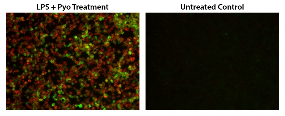
Fluorescence images of simultaneous detection of intracellular nitric oxide (NO) and total ROS in RAW 264.7 macrophage. Cells were co-stained with Nitrixyte™ Orange (Red) and Amplite® ROS Green (Green). The cells were then treated with or without 20 µg/mL of lipopolysaccharide (LPS), 1 mM L-arginine (L-Arg) and 50 µM Pyocyanin (Pyo) at 37°C for 16 hours. The fluorescence signals were measured using fluorescence microscope equipped with TRITC (Nitrixyte™ Orange, Red) and FITC (Amplite® ROS Green, Green) filter sets, simultaneously.
Detection of ROS in Jurkat cells with Cell Meter&trade; Fluorimetric Intracellular Total ROS Activity Assay Kit. Jurkat cells were seeded on the same day at 300,000 cells/100&micro;L/well in a Costar black wall/clear bottom 96-well plate. The ROS assay loading solution (100 &micro;L/well) was added and incubated in a 5% CO2, 37 &deg;C incubator for 1 hour. And then the cells were treated with 1mM, 0.1mM H<sub>2</sub>O<sub>2</sub> or without H<sub>2</sub>O<sub>2</sub> for 30 minutes. The fluorescence signal was monitored at Ex/Em = 490/525 nm (cutoff at 515 nm) with bottom read mode using FlexStation (Molecular Devices).