logo
AAT Bioquest

Cell Meter™ Cell Viability Assay Kit *NIR Fluorescence Optimized for Fluorescence Microplate Reader*

Our Cell Meter™ assay kits are a set of tools for monitoring cell viability. There are a variety of parameters that can be used for monitoring cell viability. This kit uses a proprietary dye that gets enhanced fluorescence upon entering into live cells. The dye is a hydrophobic compound that easily permeates intact live cells. The hydrolysis of the weakly fluorescent substrate by intracellular esterases generates a strongly fluorescent hydrophilic product that is well-retained in the cell cytoplasm. The esterase activity is proportional to the number of viable cells, and thus directly related to the fluorescence intensity of the product generated from the esterase-catalyzed hydrolysis of the fluorogenic substrate. Cells grown in black-walled plates can be stained and quantified in less than two hours. The assay is more robust than the tetrazolium salt or Alarmar Blue™-based assays. It can be readily adapted for high-throughput assays in a wide variety of fluorescence platforms such as microplate assays, immunocytochemistry and flow cytometry. It is useful for a variety of studies, including cell adhesion, chemotaxis, multidrug resistance, cell viability, apoptosis and cytotoxicity. The kit provides all the essential components with an optimized cell-labeling protocol. It is suitable for proliferating and non-proliferating cells, and can be used for both suspension and adherent cells. Using 100 uL of reagents per well in a 96-well format, this kit provides sufficient reagents to perform 200 assays. Using 25 uL of reagents per well in a 384-well format, this kit provides sufficient reagents to perform 800 assays.
Example protocol

AT A GLANCE

Protocol summary

  1. Prepare cells with test compounds
  2. Remove the medium
  3. Add CytoCalcein™ NIR working solution (100 µL/well/96-well plate or 25 µL/well/384-well plate)
  4. Incubate at room temperature or 37°C for 1 hr
  5. Read fluorescence intensity (bottom read mode) at Ex/Em = 635/670 nm (Cutoff = 665 nm) 

Important notes
Thaw one of each kit component at room temperature before starting the experiment.

PREPARATION OF STOCK SOLUTION

Unless otherwise noted, all unused stock solutions should be divided into single-use aliquots and stored at -20 °C after preparation. Avoid repeated freeze-thaw cycles.

1. CytoCalcein™ NIR stock solution:
Add 20 µL of DMSO (Component B) into the vial of CytoCalcein™ NIR (Component A) and mix them well to make CytoCalcein™ NIR stock solution. Note: 20 µL of CytoCalcein™ NIR stock solution is enough for one plate. Protect from light. For storage, seal tubes tightly.

PREPARATION OF WORKING SOLUTION

Add 20 µL of CytoCalcein™ NIR stock solution into the bottle of Assay Buffer (10 mL, Component C) and mix well to make CytoCalcein™ NIR working solution. Note: This CytoCalcein™ NIR working solution is for one cell plate and stable for at least 2 hours at room temperature.

SAMPLE EXPERIMENTAL PROTOCOL

Cells Preparation:

Plate 100 to 100,000×10 cells per well in a tissue culture microplate with black wall and clear bottom. Add test compounds into the cells and incubate for a desired period of time (such as 24, 48 or 96 hours) in a 37 °C, 5% CO2 incubator.

For blank wells (medium without the cells), add the same amount of compound buffer. The suggested total volume is 100 µL/well/96-well plate, and 25 µL/well/384-well plate. Note: Each cell line should be evaluated on the individual basis to determine the optimal cell density for proliferation or cytotoxicity induction. For proliferation assays, use fewer cells; for cytotoxicity assays, use more cells to start with.

Sample Protocol:

  1. Treat cells with test compounds as desired. Note: It is not necessary to wash cells before adding compound. However, if tested compounds are serum sensitive, growth medium and serum factors can be aspirated away before adding compounds. Add 100 µL/well (96-well plate) and 25 µL/well (384-well plate) of 1X Hank’s salt solution and 20 mM Hepes buffer (HHBS) or the buffer of your choice after aspiration. Alternatively, cells can be grown in serum-free media.

  2. Remove the medium from the cells. Note: Medium must be removed before loading CytoCalcein™ NIR working solution.

  3. Add 100 µL/well (96-well plate) or 25 µL/well (384-well plate) of CytoCalcein™ NIR working solution.

  4. Incubate plate at room temperature or 37°C for 1 hour, protected from light. Note: The appropriate incubation time depends on the individual cell type and cell concentration used. Optimize the incubation time for each experiment. For non-adherent cells, it is recommended to centrifuge cell plates at 800 rpm for 2 minutes with brake off after incubation.

  5. Monitor the fluorescence intensity with a fluorescence plate reader (bottom read mode) at Ex/Em = 635/670 nm (Cutoff = 665 nm).

Product family

Citations

View all 17 citations: Citation Explorer
Functional imaging of neuronal activity of auditory cortex by using Cal-520 in anesthetized and awake mice
Authors: Li, Jingcheng and Zhang, Jianxiong and Wang, Meng and Pan, Junxia and Chen, Xiaowei and Liao, Xiang
Journal: Biomedical Optics Express (2017): 2599--2610
NINJ2--A novel regulator of endothelial inflammation and activation
Authors: Wang, Jingjing and Fa, Jingjing and Wang, Pengyun and Jia, Xinzhen and Peng, Huixin and Chen, Jing and Wang, Yifan and Wang, Chenhui and Chen, Qiuyun and Tu, Xin and others, undefined
Journal: Cellular Signalling (2017)
Influence of hypothermia and subsequent rewarming upon leukocyte-endothelial interactions and expression of Junctional-Adhesion-Molecules A and B
Authors: Bogert, Nicolai V and Werner, Isabella and Kornberger, Angela and Meybohm, Patrick and Moritz, Anton and Keller, Till and Stock, Ulrich A and Beiras-Fern, undefined and ez, Andres
Journal: Scientific reports (2016)
Inhibition of ABC transport proteins by oil sands process affected water
Authors: Alharbi, Hattan A and Saunders, David MV and Al-Mousa, Ahmed and Alcorn, Jane and Pereira, Alberto S and Martin, Jonathan W and Giesy, John P and Wiseman, Steve B
Journal: Aquatic Toxicology (2016): 81--88
Rapid generation of collagen-based microtissues to study cell--matrix interactions
Authors: Brett, Marie-Elena and Crampton, Alex and ra L , undefined and Wood, David K
Journal: Technology (2016): 1--8

References

View all 84 references: Citation Explorer
Functional evidence that the self-renewal gene NANOG regulates esophageal squamous cancer development
Authors: Li, Deng and Xiang, Xiaocong and Yang, Fei and Xiao, Dongqin and Liu, Kang and Chen, Zhu and Zhang, Ruolan and Feng, Gang
Journal: Biochemical and Biophysical Research Communications (2017)
Localized functional chemical stimulation of TE 671 cells cultured on nanoporous membrane by calcein and acetylcholine
Authors: Zibek S, Stett A, Koltay P, Hu M, Zengerle R, Nisch W, Stelzle M.
Journal: Biophys J. (2006)
A vaccination and challenge model using calcein marked fish
Authors: Klesius PH, Evans JJ, Shoemaker CA, Pasnik DJ.
Journal: Fish Shellfish Immunol (2006): 20
Novel fluorescence assay using calcein-AM for the determination of human erythrocyte viability and aging
Authors: Bratosin D, Mitrofan L, Palii C, Estaquier J, Montreuil J.
Journal: Cytometry A (2005): 78
Cytotoxic effects of 100 reference compounds on Hep G2 and HeLa cells and of 60 compounds on ECC-1 and CHO cells. I mechanistic assays on ROS, glutathione depletion and calcein uptake
Authors: Schoonen WG, Westerink WM, de Roos JA, Debiton E.
Journal: Toxicol In Vitro (2005): 505
Page updated on November 21, 2024

Ordering information

Price
Unit size
Catalog Number22787
Quantity
Add to cart

Additional ordering information

Telephone1-800-990-8053
Fax1-800-609-2943
Emailsales@aatbio.com
InternationalSee distributors
Bulk requestInquire
Custom sizeInquire
Technical SupportContact us
Purchase orderSend to sales@aatbio.com
ShippingStandard overnight for United States, inquire for international
Request quotation

Storage, safety and handling

H-phraseH303, H313, H333
Hazard symbolXN
Intended useResearch Use Only (RUO)
R-phraseR20, R21, R22
UNSPSC12352200

Platform

Fluorescence microplate reader

Excitation635 nm
Emission670 nm
Cutoff665 nm
Recommended plateBlack wall, clear bottom
Instrument specification(s)Bottom read mode

Components

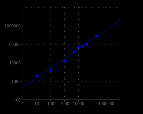
CHO-K1 cell number response was measured with Cell Meter™ Cell Viability Assay Kit. CHO-K1 cells at 0 to 50,000 cells/well/100 µL were seeded overnight in a Costar black wall/clear bottom 96-well plate. The cells were incubated with 100 µL/well of CytoCalcein™ NIR dye-working solution for 1 hour at room temperature. The fluorescence intensity was measured at Ex/Em = 635/670 nm (Cutoff = 665 nm) with FlexStation™ microplate reader (Molecular Devices).