logo
AAT Bioquest

Cell Explorer™ Live Cell Labeling Kit *Red Fluorescence*

Our Cell Explorer™ fluorescence imaging kits are a set of tools for labeling cells for fluorescence microscopic investigations of cellular functions. The effective labeling of cells provides a powerful method for studying cellular events in a spatial and temporal context. This particular kit is designed to uniformly label live cells in red fluorescence. The kit uses a proprietary non-fluorescent dye that becomes strongly fluorescence upon entering into live cells. The dye is a hydrophobic compound that easily permeates intact live cells. The hydrolysis of the non-fluorescent substrate by intracellular esterases generates a strongly red fluorescent hydrophilic product that is well-retained in the cell cytoplasm. Cells grown in black-walled plates can be stained and quantified in less than two hours. The assay is more robust than the tetrazolium salt or Alarmar Blue™-based assays. It can be readily adapted for high-throughput assays in a wide variety of fluorescence platforms such as microplate assays, immunocytochemistry and flow cytometry. It is useful in a variety of studies, including cell adhesion, chemotaxis, multidrug resistance, cell viability, apoptosis and cytotoxicity. The kit provides all the essential components with an optimized cell-labeling protocol.
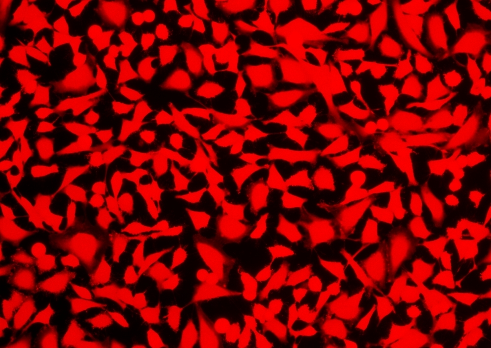
Example protocol

AT A GLANCE

Protocol Summary
  1. Prepare cells in growth medium
  2. Remove the medium
  3. Add Calcein Deep Red™ working solution (100 µL/well for 96-well plates or 25 µL/well for 384-well plates)

  4. Incubate cells at 37°C for 30 minutes to 2 hours
  5. Wash the cells
  6. Examine the specimen under under fluorescence microscope with Cy5 filter (Ex/Em = 646/660 nm)
Important Note

Thaw all the components at room temperature before starting the experiment.

CELL PREPARATION

For guidelines on cell sample preparation, please visit https://www.aatbio.com/resources/guides/cell-sample-preparation.html

PREPARATION OF STOCK SOLUTIONS

Unless otherwise noted, all unused stock solutions should be divided into single-use aliquots and stored at -20 °C after preparation. Avoid repeated freeze-thaw cycles

Calcein Deep Red™ stock solution

Add 20 µL of DMSO into the vial of Calcein Deep Red™ (Component A) and mix well to make Calcein Deep Red™ stock solution. Note: 20 µL of Calcein Deep Red™ stock solution is enough for 1 plate. Note: Unused Calcein Deep Red™ stock solution can be aliquoted and stored at < -20 °C for 2 weeks if the tubes are sealed tightly. Avoid repeated freeze-thaw cycles and protect from light.

PREPARATION OF WORKING SOLUTION

Add 20 µL of Calcein Deep Red™ stock solution into 10 mL of HHBS (Component B) and mix well to make Calcein Deep Red™ working solution. Protect from light.

SAMPLE EXPERIMENTAL PROTOCOL

  1. Remove the growth medium from the cell plates. Note: It is important to remove the growth medium in order to minimize the background fluorescence and increase the signal to background ratio.
  2. Add 100 µL/well (96-well plate) or 25 µL/well (384-well plate) Calcein Deep Red™ working solution into the cell plate.

  3. Incubate the cells in a 37°C, 5% CO2 incubator for 30 minutes to 2 hours.
  4. Remove the Calcein Deep Red™ working solution from the cells.

  5. Wash the cells with HHBS (Component B) for 2 to 3 times, and replace with HHBS.
  6. Image the cells using a fluorescence microscope with Cy5 filter (Ex/Em = 646/660 nm).

Spectrum

Product family

Citations

View all 4 citations: Citation Explorer
Autophagy proteins are not universally required for phagosome maturation
Authors: Cemma, Marija and Grinstein, Sergio and Brumell, John H
Journal: Autophagy (2016): 1440--1446
Size control and biological properties of monodispersed mesoporous bioactive glass sub-micron spheres
Authors: Hu, Qing and Li, Yuli and Miao, Guohou and Zhao, Naru and Chen, Xiaofeng
Journal: Rsc Advances (2014): 22678--22687
Differential detection of tumor cells using a combination of cell rolling, multivalent binding, and multiple antibodies
Authors: Myung, Ja Hye and Gajjar, Khyati A and Chen, Jihua and Molokie, Robert E and Hong, Seungpyo
Journal: Analytical chemistry (2014): 6088--6094
Versatile fabrication of nanoscale sol--gel bioactive glass particles for efficient bone tissue regeneration
Authors: Lei, Bo and Chen, Xiaofeng and Han, Xue and Zhou, Jiaan
Journal: Journal of Materials Chemistry (2012): 16906--16913

References

View all 26 references: Citation Explorer
Requirements, features, and performance of high content screening platforms
Authors: Gough AH, Johnston PA.
Journal: Methods Mol Biol (2007): 41
A pharmaceutical company user's perspective on the potential of high content screening in drug discovery
Authors: Hoffman AF, Garippa RJ.
Journal: Methods Mol Biol (2007): 19
Optimizing the integration of immunoreagents and fluorescent probes for multiplexed high content screening assays
Authors: Giuliano KA., undefined
Journal: Methods Mol Biol (2007): 189
Past, present, and future of high content screening and the field of cellomics
Authors: Taylor DL., undefined
Journal: Methods Mol Biol (2007): 3
G protein-coupled receptor internalization assays in the high-content screening format
Authors: Haasen D, Schnapp A, Valler MJ, Heilker R.
Journal: Methods Enzymol (2006): 121
Page updated on January 18, 2025

Ordering information

Price
Unit size
Catalog Number22609
Quantity
Add to cart

Additional ordering information

Telephone1-800-990-8053
Fax1-800-609-2943
Emailsales@aatbio.com
InternationalSee distributors
Bulk requestInquire
Custom sizeInquire
Technical SupportContact us
Purchase orderSend to sales@aatbio.com
ShippingStandard overnight for United States, inquire for international
Request quotation

Spectral properties

Excitation (nm)

643

Emission (nm)

663

Storage, safety and handling

H-phraseH303, H313, H333
Hazard symbolXN
Intended useResearch Use Only (RUO)
R-phraseR20, R21, R22
UNSPSC12352200

Platform

Fluorescence microscope

ExcitationCy5 filter set
EmissionCy5 filter set
Recommended plateBlack wall, clear bottom

Components