logo
AAT Bioquest

Cal-630™ AM

Calcium measurement is critical for numerous biological investigations. Fluorescent probes that show spectral responses upon binding calcium have enabled researchers to investigate changes in intracellular free calcium concentrations by using fluorescence microscopy, flow cytometry, fluorescence spectroscopy and fluorescence microplate readers. x-Rhod-1 is commonly used as a red fluorescent calcium indicator. However, x-Rhod-1 is only moderately fluorescent in live cells upon esterase hydrolysis, and has very small cellular calcium responses. Cal-630™ has been developed to improve x-Rhod-1 cell loading and calcium response while maintaining the spectral wavelength of x-Rhod-1, making it compatible with Texas Red® filter set. In CHO and HEK cells Cal-630™ AM has cellular calcium response that is much more sensitive than x-Rhod-1. The spectra of Cal-630 is well separated from those of FITC, Alexa Fluor® 488 and GFP, making it an ideal calcium probe for multiplexing intracellular assays with GFP cell lines or FITC/Alexa Fluor® 488 labeled antibodies.
Example protocol

PREPARATION OF STOCK SOLUTIONS

Unless otherwise noted, all unused stock solutions should be divided into single-use aliquots and stored at -20 °C after preparation. Avoid repeated freeze-thaw cycles

Cal-630™ AM Stock Solution
  1. Prepare a 2 to 5 mM stock solution of Cal-630™ AM in anhydrous DMSO.

    Note: When reconstituted in DMSO, Cal-630™ AM is a clear, colorless solution.

PREPARATION OF WORKING SOLUTION

Cal-630™ AM Working Solution
  1. On the day of the experiment, either dissolve Cal-630™ AM in DMSO or thaw an aliquot of the indicator stock solution to room temperature.

  2. Prepare a 2 to 20 µM Cal-630™ AM working solution in a buffer of your choice (e.g., Hanks and Hepes buffer) with 0.04% Pluronic® F-127. For most cell lines, Cal-630™ AM at a final concentration of 4-5 μM is recommended. The exact concentration of indicators required for cell loading must be determined empirically.

    Note: The nonionic detergent Pluronic® F-127 is sometimes used to increase the aqueous solubility of Cal-630™ AM. A variety of Pluronic® F-127 solutions can be purchased from AAT Bioquest.

    Note: If your cells contain organic anion-transporters, probenecid (1-2 mM) may be added to the dye working solution (final in well concentration will be 0.5-1 mM) to reduce leakage of the de-esterified indicators. A variety of ReadiUse™ Probenecid products, including water-soluble, sodium salt, and stabilized solutions, can be purchased from AAT Bioquest.

SAMPLE EXPERIMENTAL PROTOCOL

Following is our recommended protocol for loading AM esters into live cells. This protocol only provides a guideline and should be modified according to your specific needs.

  1. Prepare cells in growth medium overnight.
  2. On the next day, add 1X Cal-630™ AM working solution to your cell plate.

    Note: If your compound(s) interfere with the serum, replace the growth medium with fresh HHBS buffer before dye-loading.

  3. Incubate the dye-loaded plate in a cell incubator at 37 °C for 30 to 60 minutes.

    Note: Incubating the dye for longer than 2 hours can improve signal intensities in certain cell lines.

  4. Replace the dye working solution with HHBS or buffer of your choice (containing an anion transporter inhibitor, such as 1 mM probenecid, if applicable) to remove any excess probes.
  5. Add the stimulant as desired and simultaneously measure fluorescence using either a fluorescence microscope equipped with a Texas Red filter set or a fluorescence plate reader containing a programmable liquid handling system such as an FDSS, FLIPR, or FlexStation, at Ex/Em = 600/640 nm cutoff 630 nm.

Calculators

Common stock solution preparation

Table 1. Volume of DMSO needed to reconstitute specific mass of Cal-630™ AM to given concentration. Note that volume is only for preparing stock solution. Refer to sample experimental protocol for appropriate experimental/physiological buffers.

0.1 mg0.5 mg1 mg5 mg10 mg
1 mM77.949 µL389.745 µL779.49 µL3.897 mL7.795 mL
5 mM15.59 µL77.949 µL155.898 µL779.49 µL1.559 mL
10 mM7.795 µL38.975 µL77.949 µL389.745 µL779.49 µL

Molarity calculator

Enter any two values (mass, volume, concentration) to calculate the third.

Mass (Calculate)Molecular weightVolume (Calculate)Concentration (Calculate)Moles
/=x=

Spectrum

Product family

NameExcitation (nm)Emission (nm)Quantum yield
Cal-590™ AM5745880.621
Cal-520®, AM4925150.751
Cal-520FF™, AM4925150.751
Cal-520N™, AM4925150.751
Calbryte™ 630 AM607624-
Cal-500™ AM3884820.481
Cal-520ER™ AM492515-

Citations

View all 11 citations: Citation Explorer
Inspiration and inflammation in the breathing brainstem
Authors: Reising, Jan Philipp
Journal: (2024)
Triple-Decker Sandwich Cultures of Intestinal Organoids for Long-Term Live Imaging, Uniform Perturbation, and Statistical Sampling
Authors: Cambra, Hailey M and Tallapragada, Naren P and Mannam, Prabhath and Breault, David T and Klein, Allon M
Journal: Current protocols (2022): e330
Tetraspanin-7 regulation of L-type voltage-dependent calcium channels controls pancreatic $\beta$-cell insulin secretion
Authors: Dickerson, Matthew T and Dadi, Prasanna K and Butterworth, Regan B and Nakhe, Arya Y and Graff, Sarah M and Zaborska, Karolina E and Schaub, Charles M and Jacobson, David A
Journal: The Journal of Physiology (2020): 4887--4905
Development of a deep two-photon calcium imaging method for the analysis of cortical processing in the mammalian brain
Authors: Birkner, Antje
Journal: (2019)

References

View all 8 references: Citation Explorer
Protein kinase C and myocardial calcium handling during ischemia and reperfusion: lessons learned using Rhod-2 spectrofluorometry
Authors: Stamm C, del Nido PJ.
Journal: Thorac Cardiovasc Surg (2004): 127
Cytosolic calcium in the ischemic rabbit heart: assessment by pH- and temperature-adjusted rhod-2 spectrofluorometry
Authors: Stamm C, Friehs I, Choi YH, Zurakowski D, McGowan FX, del Nido PJ.
Journal: Cardiovasc Res (2003): 695
Calcium measurements in perfused mouse heart: quantitating fluorescence and absorbance of Rhod-2 by application of photon migration theory
Authors: Du C, MacGowan GA, Farkas DL, Koretsky AP.
Journal: Biophys J (2001): 549
Calibration of the calcium dissociation constant of Rhod(2)in the perfused mouse heart using manganese quenching
Authors: Du C, MacGowan GA, Farkas DL, Koretsky AP.
Journal: Cell Calcium (2001): 217
Changes in mitochondrial Ca2+ detected with Rhod-2 in single frog and mouse skeletal muscle fibres during and after repeated tetanic contractions
Authors: Lannergren J, Westerblad H, Bruton JD.
Journal: J Muscle Res Cell Motil (2001): 265
Page updated on January 22, 2025

Ordering information

Price
AvailabilityIn stock
Unit size
5x50 ug
10x50 ug
1 mg
Catalog Number
205302053120532
Quantity
Add to cart

Additional ordering information

Telephone1-800-990-8053
Fax1-800-609-2943
Emailsales@aatbio.com
InternationalSee distributors
Bulk requestInquire
Custom sizeInquire
Technical SupportContact us
Purchase orderSend to sales@aatbio.com
ShippingStandard overnight for United States, inquire for international
Request quotation

Physical properties

Dissociation constant (Kd, nM)792

Molecular weight

1282.89

Solvent

DMSO

Spectral properties

Excitation (nm)

609

Emission (nm)

626

Quantum yield

0.371

Storage, safety and handling

H-phraseH303, H313, H333
Hazard symbolXN
Intended useResearch Use Only (RUO)
R-phraseR20, R21, R22

Storage

Freeze (< -15 °C); Minimize light exposure
UNSPSC12352200

Platform

Fluorescence microscope

ExcitationTexas Red
EmissionTexas Red
Recommended plateBlack wall, clear bottom

Fluorescence microplate reader

Excitation600
Emission640
Cutoff630
Recommended plateBlack wall, clear bottom
Instrument specification(s)Bottom read mode, Programmable liquid handling
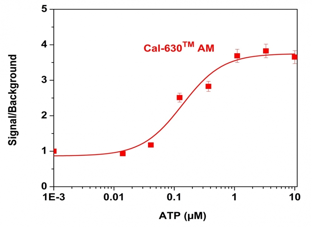
ATP-stimulated calcium responses of endogenous P2Y receptor in CHO-K1 cells incubated with Cal-630&trade; AM (red curve). CHO-K1 cells were seeded overnight at 50,000 cells per 100 uL per well in a Costar black wall/clear bottom 96-well plate. 100 uL of 5 &micro;M Cal-630 &trade; AM in HHBS (with 1.0 mM probenecid) was added into the cells and incubated at 37 &deg;C for 1 hour. ATP (50 uL/well) was added using FlexSation to achieve the final indicated concentrations.
Response of endogenous P2Y receptor to ATP in CHO-K cells detected with Cal-630 &trade; AM.&nbsp; CHO-K1 cells were seeded overnight at 50,000 cells per 100 &micro;L per well in a Costar black wall/clear bottom 96-well plate. 100 uL of 5 uM Cal-630 &trade; AM in HHBS (with 1.0 mM probenecid) was added into the cells and incubated at 37 &deg;C for 1 hour.&nbsp; Images were recorded with a fluorescence microscope (Olympus IX71) before and after adding 10 uM ATP (final in the well) using Texas Red Channel.