logo
AAT Bioquest

Buccutite™ ALP (Alkaline Phosphatase) Antibody Conjugation Kit *Optimized for Labeling 100 ug Protein*

Protein-protein conjugations are commonly performed with a bifunctional linker (such as SMCC) having different reactivity at each end for linking two different proteins. One end of the crosslinker reacts (via NHS ester) with amines (-NH2) of lysine and N-terminus, and the other end reacts (via maleimide) with the thiol groups (-SH) of cysteine. However, SMCC-modified protein is extremely unstable and often self-reactive since proteins contain multiple amine and thiol groups that cause significant amount of crosslinkings. In addition, it is quite difficult and tedious to quantify the number of maleimide groups in a protein. Buccutite™ ALP (Alkaline Phosphatase) Antibody Conjugation Kit is designed to rapidly prepare ALP conjugates with pre-activated ALP. The kit is optimized for labeling 100 ug protein. The Buccutite™ FOL-activated ALP readily reacts with Buccutite™ MTA-modified antibody under extremely mild neutral conditions without any catalyst required. Compared with commonly used SMCC and other similar technologies, Buccutite™ bioconjugation system is much more robust and easier to use. It enables faster and quantitative conjugation of biomolecules with higher efficiency and yields.
Example protocol

AT A GLANCE

Protocol summary
  1. Add 5 µL of Reaction Buffer (Component C) into antibody (100 µL)
  2. Add the antibody solution into Buccutite™ MTA vial (Component B) to antibody working solution
  3. Incubate the mixture at room temperature for 30 minutes
  4. Mix the antibody- Buccutite ™ MTA reaction mixture with Buccutite™ FOL-Activated ALP (Component A)
  5. Incubate at room temperature for 60 minutes 

Important
Upon receipt, store the kit at 4 °C. When stored properly, the kit should be stable for six months. Alternatively, Component B can be stored at -20 °C. Warm up all the components at room temperature and centrifuge the vials briefly before opening. Immediately prepare the required solutions before starting your conjugation. The following SOP is an example for labeling 100 µg goat anti-mouse IgG antibody.

PREPARATION OF WORKING SOLUTION

Antibody working solution
For labeling 100 µg antibody (assuming the target antibody concentration is 1 mg/mL), mix 5 µL (5% of the total reaction volume) of Reaction Buffer (Component C) with 100 µL of the target antibody solution.
Note     If you have a different concentration, adjust the antibody volume accordingly to make ~100 µg antibody /100 uL available for your labeling reaction.
Note     The antibody should be dissolved in 1X phosphate buffered saline (PBS), pH 7.2-7.4. If the antibody is dissolved in glycine buffer, it must be dialyzed against 1X PBS, pH 7.2-7.4, or use 10 kDa Filter (Cat. # 60502 from AAT Bioquest) to remove free amines or ammonium salts (such as ammonium sulfate and ammonium acetate).
Note     Impure antibodies or antibodies stabilized with bovine serum albumin (BSA) or gelatin will not be labeled well.
Note     The antibody –Buccutite™ MTA reaction efficiency is significantly reduced if the antibody concentration is less than 1 mg/mL. For optimal labeling efficiency, the final antibody concentration range of 1-10 mg/mL is recommended.

SAMPLE EXPERIMENTAL PROTOCOL

Run Antibody-Buccutite™ MTA reaction
  1. Add the antibody solution directly into the vial of Buccutite ™ MTA (Component B), and mix them well by repeatedly pipetting for a few times or vortex the vial for a few seconds.
  2. Keep the antibody- Buccutite ™ MTA reaction mixture at room temperature for 30 minutes.
    Note     The antibody-Buccutite™ MTA reaction mixture can be rotated or shaken for longer time if desired. 

Make antibody-ALP conjugation
  1. Add antibody-Buccutite™ MTA reaction mixture to the vial of Buccutite™ FOL-Activated ALP (Component A), mix well and incubate the mixture at room temperature for 1 hour.
    Note     The antibody-ALP reaction mixture can be incubated for longer time if desired.
  2. The antibody-ALP conjugate is now ready to use.
    Note     For immediate use, the antibody-ALP conjugate needs to be diluted with the buffer of your choice.
  3. The concentration of the conjugate can be calculated as follows: Antibody Concentration (µg/uL) = 100 µg (total amount of antibody)/(100 µL + 5 µL) = 0.95 µg/µL 

Storage of Antibody-ALP Conjugate
The antibody conjugate-ALP conjugate should be stored in the presence of a carrier protein (e.g., 0.1% bovine serum albumin) at 4 °C and kept from light for two months.
Page updated on November 20, 2024

Ordering information

Price
Unit size
Catalog Number5513
Quantity
Add to cart

Additional ordering information

Telephone1-800-990-8053
Fax1-800-609-2943
Emailsales@aatbio.com
InternationalSee distributors
Bulk requestInquire
Custom sizeInquire
Technical SupportContact us
Purchase orderSend to sales@aatbio.com
ShippingStandard overnight for United States, inquire for international
Request quotation

Storage, safety and handling

H-phraseH303, H313, H333
Hazard symbolXN
Intended useResearch Use Only (RUO)
R-phraseR20, R21, R22
UNSPSC12171501

Components

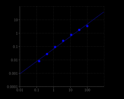
Mouse IgG dose curve was detected using the ALP- goat-anti-mouse IgG conjugate prepared with Buccutite™ ALP (Alkaline Phosphatase) Antibody Conjugation Kit. 3-fold  serial diluted mouse IgG was coated on a 96-well plate, and  100uL GAM IgG-ALP conjugate (1ug/ml) was added to each well followed the standard ELISA method. pNPP substrate solution was used to detect the immobilized mouse IgG with 30 min incubation and read at 400 nm.