logo
AAT Bioquest

Amylite™ Red

Amylite™ Red staining on healthy and Alzheimer's disease brain tissue with DAPI counterstain.
Ordering information
Price
Catalog Number
Unit Size
Quantity
Add to cart
Additional ordering information
Telephone1-800-990-8053
Fax1-800-609-2943
Emailsales@aatbio.com
InternationalSee distributors
Bulk requestInquire
Custom sizeInquire
ShippingStandard overnight for United States, inquire for international
Request quotation
Physical properties
Molecular weight391.23
SolventDMSO
Spectral properties
Excitation (nm)485
Emission (nm)671
Storage, safety and handling
H-phraseH303, H313, H333
Hazard symbolXN
Intended useResearch Use Only (RUO)
R-phraseR20, R21, R22
StorageFreeze (< -15 °C); Minimize light exposure

OverviewpdfSDSpdfProtocol


Molecular weight
391.23
Excitation (nm)
485
Emission (nm)
671
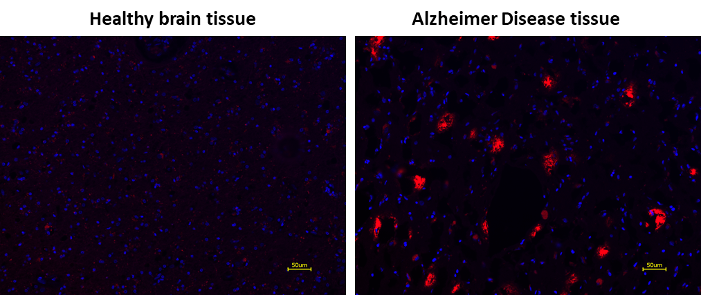
The accumulation of insoluble amyloid plaques in the nerve tissue is widely considered to contribute to the development of neurodegenerative human diseases, such as Alzheimer’s disease. The investigation of the beta-amyloid formation is one of the most important tasks to find an effective way to inhibit the accumulation of insoluble amyloid plaques in the nerve tissue. However, the lack of effective imaging tools severely limited this research. Staining of nerve tissues with Congo Red dye is one of the most common tools for monitoring the formation of the amyloid aggregates. Congo Red has a few limitations such as its low sensitivity, significant toxicity and carcinogenicity. AAT Bioquest developed Amylite™ Red stain to address these limitations. Amylite™ Red is designed to label amyloid plaques in paraffin-embedded or freshly cut frozen tissue sections via a simple mix and read step. Amylite™ Red staining can be completed within 30 minutes with desired specificity. The staining process does not require antibodies. It is much more efficient and cost effective than the anti-amyloid antibody-based fluorescence imaging. Amylite™ Red imaging reagent is compatible with other fluorophores, such as DAPI, Hoechst and ethidium bromide, as well as fluorescent-labelled antibodies with emission spectra in the blue or green emission range, such as GFP.

Platform


Fluorescence microscope

Excitation510 nm
Emission660 nm
Recommended plateBlack wall/clear bottom

Example protocol


AT A GLANCE

Important Note

Before using Amylite™ Red for the first time, thaw the labeling dye at room temperature and briefly centrifuge to gather the dried pellet.

Protocol Summary
  1. Prepare the tissue slides.

  2. Add the Amylite™ Red working solution to the tissue sections.

  3. Incubate at room temperature for 3 to 5 minutes.

  4. Wash slides with 40% ethanol.

  5. Conduct counterstaining with DAPI as needed.

  6. Mount the samples with a suitable mounting medium. Then, monitor the staining using a fluorescence microscope with a Cy3 filter set.

PREPARATION OF STOCK SOLUTIONS

Unless otherwise noted, all unused stock solutions should be divided into single-use aliquots and stored at -20 °C after preparation. Avoid repeated freeze-thaw cycles

Amylite™ Red Stock Solution (100X)
  1. To prepare a 100X Amylite™ Red stock solution, add 100 μL of DMSO to the Amylite™ Red vial.

    Note: Prepare a single aliquot of any unused Amylite™ Red stock solution and store it at ≤-20°C, protected from light. Avoid freeze/thaw cycles.

PREPARATION OF WORKING SOLUTION

Amylite™ Red Working Solution
  1. Prepare the Amylite™ Red working solution by adding 10 μL of Amylite™ Red stock solution to 1 mL of 50% ethanol solution. Protect the Amylite™ Red working solution from light by covering it with foil or placing it in a dark location.

    Note: For optimal results, use this solution within a few hours of preparation.

    Note: 1 mL of the working solution is sufficient for 10 tests.

SAMPLE EXPERIMENTAL PROTOCOL

Tissue Preparation (Deparaffinization and Rehydration):
  1. Wash the slides with Xylene for 3 minutes.

  2. Wash the slides with 100% Ethanol for 1 minute.

  3. Wash the slides with water for 2 minutes.

Tissue Staining
  1. Add 100 μL of Amylite™ Red working solution to the tissue sections.

    Note: Ensure sufficient solution is added to thoroughly cover the tissue slides.

  2. Incubate the slides at room temperature for 3 to 5 minutes.

    Note: The incubation time can be adjusted for optimal results.

  3. Wash the tissue slides twice with 40% ethanol, for 5 minutes each time.

  4. Use a suitable counterstain, such as DAPI, if needed.

  5. Apply the mounting medium to the slides and allow them to dry.

  6. Observe the slides using a fluorescence microscope equipped with a Cy3 filter set.

Calculators


Common stock solution preparation

Table 1. Volume of DMSO needed to reconstitute specific mass of Amylite™ Red to given concentration. Note that volume is only for preparing stock solution. Refer to sample experimental protocol for appropriate experimental/physiological buffers.

0.1 mg0.5 mg1 mg5 mg10 mg
1 mM255.604 µL1.278 mL2.556 mL12.78 mL25.56 mL
5 mM51.121 µL255.604 µL511.208 µL2.556 mL5.112 mL
10 mM25.56 µL127.802 µL255.604 µL1.278 mL2.556 mL

Molarity calculator

Enter any two values (mass, volume, concentration) to calculate the third.

Mass (Calculate)Molecular weightVolume (Calculate)Concentration (Calculate)Moles
/=x=

Spectrum


Open in Advanced Spectrum Viewer
spectrum

Spectral properties

Excitation (nm)485
Emission (nm)671

Product Family


NameExcitation (nm)Emission (nm)
Amplite® Red571584
Nile Red *CAS 7385-67-3*559635
LysoBrite™ Red576596

Images


References


View all 50 references: Citation Explorer
A role of dentate gyrus mechanistic target of rapamycin activation in epileptogenesis in a mouse model of posttraumatic epilepsy.
Authors: Guo, Dongjun and Han, Lirong and Godale, Christin M and Rensing, Nicholas R and Danzer, Steve C and Wong, Michael
Journal: Epilepsia (2024)
LAMC1 attenuates neuronal apoptosis via FAK/PI3K/AKT signaling pathway after subarachnoid hemorrhage.
Authors: Wu, Qiaowei and Yuan, Kaikun and Yao, Yanting and Yao, Jinbiao and Shao, Jiang and Meng, Yuxiao and Wu, Pei and Shi, Huaizhang
Journal: Experimental neurology (2024): 114776
MiR129-5p-loaded exosomes suppress seizure-associated neurodegeneration in status epilepticus model mice by inhibiting HMGB1/TLR4-mediated neuroinflammation.
Authors: Liu, Tengfei and Liu, Haiyan and Xue, Siyi and Xiao, Lijie and Xu, Jing and Tong, Shuyan and Wei, Xiu'e
Journal: Molecular biology reports (2024): 292
Propofol and Dexmedetomidine Ameliorate Endotoxemia-Associated Encephalopathy via Inhibiting Ferroptosis.
Authors: Zhou, Ye and Yang, Yangliang and Yi, Liang and Pan, Mengzhi and Tang, Weiqing and Duan, Hongwei
Journal: Drug design, development and therapy (2024): 1349-1368
Compartmental neuronal degeneration in the ventral striatum induced by status epilepticus in young rats' brain in comparison with adults.
Authors: Al-Redouan, Azzat and Salaj, Martin and Kubova, Hana and Druga, Rastislav
Journal: International journal of developmental neuroscience : the official journal of the International Society for Developmental Neuros (2024): 328-341
Evaluation of the anticonvulsant and neuroprotective effect of intracerebral administration of growth hormone in rats.
Authors: Zamora-Bello, I and Martínez, A and Beltrán-Parrazal, L and Santiago-Roque, I and Juárez-Aguilar, E and López-Meraz, M L
Journal: Neurologia (2024): 1-9
Attenuation of Nerve Agent Induced Neurodegenerative and Neuroinflammatory Changes in Rats with New Combination Treatment of Galantamine, Atropine and Midazolam.
Authors: Singh, Naveen and Golime, RamaRao and Kumar, Abdhesh and Roy, Tuhin
Journal: Molecular neurobiology (2024)
Mas receptor activation facilitates innate hematoma resolution and neurological recovery after hemorrhagic stroke in mice.
Authors: Deng, Xiangyang and Ren, Junwei and Chen, Kezhu and Zhang, Jin and Zhang, Quan and Zeng, Jun and Li, Tianwen and Tang, Qisheng and Lin, Jian and Zhu, Jianhong
Journal: Journal of neuroinflammation (2024): 106
Electroacupuncture Downregulating Neuronal Ferroptosis in MCAO/R Rats by Activating Nrf2/SLC7A11/GPX4 Axis.
Authors: Zhu, Wei and Dong, Jianjian and Han, Yongsheng
Journal: Neurochemical research (2024)
Intranasal administration of recombinant prosaposin attenuates neuronal apoptosis through GPR37/PI3K/Akt/ASK1 pathway in MCAO rats.
Authors: Yu, Jing and Li, Jinlan and Matei, Nathanael and Wang, Wenna and Tang, Lihui and Pang, Jinwei and Li, Xue and Fang, Lili and Tang, Jiping and Zhang, John H and Yan, Min
Journal: Experimental neurology (2024): 114656