Amplite® Universal Fluorimetric Protease Activity Assay Kit *Red Fluorescence*
Example protocol
AT A GLANCE
Protocol summary
Measuring protease activity in the test sample (Protocol A)
- Prepare Protease Substrate working solution (50 µL)
- Add substrate control, positive control or test samples (50 µL)
- Skip incubation for kinetic reading or incubate 30 minutes - 1 hour for end point reading
- Monitor fluorescence intensity at Ex/Em = 540/590 nm (Cutoff = 570 nm)
Protocol summary
Screening protease inhibitors using a purified enzyme (Protocol B)
- Prepare Protease Substrate working solution (10 µL)
- Add substrate control, positive control, vehicle control or test samples (90 µL)
- Skip incubation for kinetic reading or incubate 30 minutes -1 hour for end point reading
- Monitor fluorescence intensity at Ex/Em = 540/590 nm (Cutoff = 570 nm)
Important notes
Thaw all the kit components at room temperature before starting the experiment. Please choose Protocol A or Protocol B according to your needs.
PREPARATION OF WORKING SOLUTION
For Protocol A
1. Protease Substrate working solution (for Protocol A):
Dilute Protease Substrate (Component A) at 1:100 in 2X Assay Buffer (Component C) to make Protease Subtrate working solution for Protocol A. Use 50 µL/well of Protease Substrate working solution for a 96-well plate. Note: The 2X Assay Buffer (Component C) is designed for detecting the activity of chymotrypsin, trypsin, thermolysin, proteinase K, protease XIV, and human leukocyte elastase. For other proteases, please refer to Table 1 for the appropriate assay buffer formula.
2. Trypsin dilution:
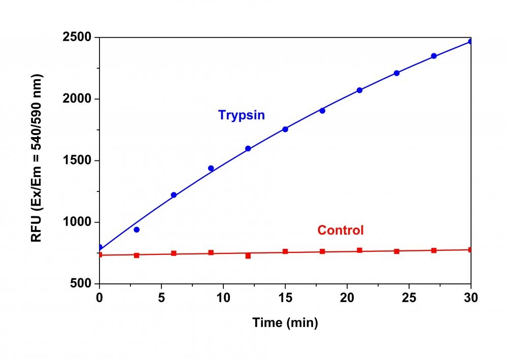
Dilute Trypsin (5 U/µL, Component B) at 1:50 in de-ionized water to get a concentration of 0.1 U/µL Trypsin dilution.
For Protocol B
1. Assay buffer (1X):
Add 5 mL of de-ionized water to 5 mL of 2X Assay Buffer (Component C) to make 1X Assay buffer.
2. Protease Substrate working solution (for Protocol B) :
Dilute Protease Substrate (Component A) at 1:20 in 1X Assay buffer to make Protease Substrabe working solution for Protocol B. Use 10 µL/well of Protease Substrate working solution for a 96-well plate. Note: The 2X Assay Buffer (Component C) is designed for detecting the activity of chymotrypsin, trypsin, thermolysin, proteinase K, protease XIV, and human leukocyte elastase. For other proteases, please refer to Table 1 for the appropriate assay buffer formula.
3. Protease dilution:
Dilute the protease in 1X assay buffer to a concentration of 500 - 1000 nM (For Trypsin 50-100 U/mL). Each well will need 10 µL of protease diluent. Prepare an appropriate amount for all the test samples and extra for the positive control and vehicle control wells.
Table 1. Assay buffer formulas for proteases. For Protocol A, 2X assay buffer is needed. For Protocol B, 1X assay buffer is needed.
Protease | 1X or 2X Assay Buffer |
Cathepsin D | 20 mM Sodium Citrate, pH 3.0 |
Papain | 20 mM sodium acetate, 20 mM cysteine, 2 mM EDTA, pH 6.5 |
PAE | 20 mM sodium phosphate, pH 8.0 |
Pepsin | 10 mM HCl, pH 2.0 |
Porcine pancreas elastase | 10 mM Tris-HCl, pH 8.8 |
Subtilisin | 20 mM potassium phosphate buffer, pH 7.6, 150 mM NaCl |
SAMPLE EXPERIMENTAL PROTOCOL
Protocol A: Measuring protease activity in test samples
Table 2A. Layout of the substrate control, positive control, and test samples in a solid black 96-well microplate. SC=Substrate Control, PC =Positive Control, TS=Test Samples.
SC | SC | ... | ... |
PC | PC | ... | ... |
TS | TS | ||
... | ... | ||
... | ... | ||
Table 3A. Reagent composition for each well. If less than 50 µL of protease-containing biological sample is used, add ddH2O to make a total volume of 50 µL.
Well | Volume | Reagent |
SC | 50 µL | De-ionized water |
PC | 50 µL | Typsin dilution |
TS | 50 µL | Protease-containing samples |
- Prepare Subtrate Control (SC), Positive Control (PC) and Test Samples (TS) according to the layout provided in Table 2A and Table 3A.
- Add 50 µL of Protease Substrate working solution (Protocol A) to all the wells in the assay plate. Mix the reagents well.
- Monitor the fluorescence increase with a fluorescence plate reader at Ex/Em = 540/590 nm (Cutoff = 570 nm).
For kinetic reading: Immediately start measuring fluorescence intensity continuously and record data every 5 minutes for 30 minutes.
For end-point reading: Incubate the reaction at a desired temperature for 30 to 60 minutes, protected from light. Then measure the fluorescence intensity.
Protocol B: Screening protease inhibitors using a purified enzyme
Table 2B. Layout of the samples in a solid black 96-well microplate. SC=Substrate Control, PC= Positive Control, VC=Vehicle Control, TS=Test Samples. It is recommended to test at least three different concentrations of each test compound. All the test samples should be done in duplicates or triplicates.
SC | SC | ... | ... |
PC | PC | ... | ... |
VC | VC | ||
TS | TS | ||
... | ... | ||
... | ... | ||
Table 3B. Reagent composition for each well. For each volume of test compound added into a well, the same volume of solvent used to deliver test compound needs to be checked for the effect of vehicle on the activity of protease.
Well | Volume | Reagent |
SC | 90 µL | Assay Buffer (1X) (90 µL) |
PC |
90 µL |
Assay Buffer (1x) (80 µL) |
VC | 90 µL |
Vehicle (X µL) |
TS | 90 µL | Test compound (X µL) Assay Buffer (1X) (80 -X µL) Protease dilution (10 µL) |
- Prepare Subtrate Control (SC), Positive Control (PC), Vehicle Control (VC) and Test Samples (TS) according to the layout provided in Table 2B and Table 3B.
- Add 10 µL of Protease Substrate working solution (Protocol B) into the wells of substrate control (PC), positive control (PC), vehicle control (VC), and test sample (TS) wells. Mix the reagents well.
- Monitor the fluorescence intensity with a fluorescence plate reader at Ex/Em = 540/590 nm (Cutoff = 570 nm).
For kinetic reading: Immediately start measuring fluorescence intensity continuously and record data every 5 minutes for 30 minutes.
For end-point reading: Incubate the reaction at a desired temperature for 30 to 60 minutes, protected from light. Then measure the fluorescence intensity.
Spectrum
Product family
Name | Excitation (nm) | Emission (nm) | Extinction coefficient (cm -1 M -1) | Quantum yield | Correction Factor (280 nm) |
Amplite® Universal Fluorimetric Protease Activity Assay Kit *Green Fluorescence* | 491 | 516 | 73000 | 0.92 | 0.35 |
Citations
Authors: Altin-Yavuzarslan, Gokce and Drake, Kinsey and Yuan, Shuo-Fu and Brooks, Sierra M and Kwa, Eng and Alper, Hal S and Nelson, Alshakim
Journal: Matter (2024)
Authors: Ullah, Inam and Uddin, Shahab and Zhao, Longhe and Wang, Xin and Li, Hongyu
Journal: Experimental Brain Research (2024): 1--16
Authors: Sugianto, Widianti and Altin-Yavuzarslan, Gokce and Tickman, Benjamin I and Kiattisewee, Cholpisit and Yuan, Shuo-Fu and Brooks, Sierra M and Wong, Jitkanya and Alper, Hal S and Nelson, Alshakim and Carothers, James M
Journal: Materials Today Bio (2023): 100677
Authors: Seiser, S and Cerbu, D and Gallhofer, A and Matiasek, J and Elbe-B{\"u}rger, A
Journal: Scientific reports (2022): 1--9
Authors: Ueki, Shigeharu and Konno, Yasunori and Takeda, Masahide and Moritoki, Yuki and Hirokawa, Makoto and Matsuwaki, Yoshinori and Honda, Kohei and Ohta, Nobuo and Yamamoto, Shiori and Takagi, Yuri and others, undefined
Journal: Journal of Allergy and Clinical Immunology (2016): 258--267
References
Authors: Vineyard D, Zhang X, Lee I.
Journal: Biochemistry (2006): 11432
Authors: Yadav SC, P and e M, Jagannadham MV.
Journal: Phytochemistry (2006): 1414
Authors: Frohbieter KA, Ismail B, Nielsen SS, Hayes KD.
Journal: J Dairy Sci (2005): 3392
Authors: Chauhan V, Sheikh AM, Chauhan A, Spivack WD, Fenko MD, Malik MN.
Journal: J Alzheimers Dis (2005): 37
Authors: Cilenti L, Lee Y, Hess S, Srinivasula S, Park KM, Junqueira D, Davis H, Bonventre JV, Alnemri ES, Zervos AS.
Journal: J Biol Chem (2003): 11489