logo
AAT Bioquest

Amplite® Universal Fluorimetric Kinase Assay Kit *Red Fluorescence*

Most of commercial protein kinase assay kits are either based on monitoring of phosphopeptide formation or ATP deletion. For the kinase assay kits that are based on detection of phosphopeptides one has to spend time and efforts to identify an optimized peptide substrate while the ATP depletion method suffers various interferences due to the use of luciferase that are inhibited or activated by various biological compounds. The Amplite® Universal Kinase Assay Kit is based on the monitoring of ADP formation, which is directly proportional to enzyme phoshphotransferase activity and is measured fluorimetricaly. This kit provides a fast, simple, and homogeneous assay for measure kinases activities. The characteristics of its high sensitivity (<0.2 uM ADP), broad ATP tolerance (1-300 uM), non-antibody based, non-radioactive and no-wash method to detect the amount of ADP produced as a result of enzyme activity make it an ideal kit for determining kinase Michaelis-Menten kinetics and for screening and identifying kinase inhibitors. The assay can be performed in a convenient 96-well or 384-well microtiter-plate format and easily adapted to automation with no separation steps required.
Example protocol

AT A GLANCE

Protocol Summary
  1. Run kinase reaction (20 µL)
  2. Add ADP Sensor Buffer (20 µL)
  3. Add ADP Sensor working solution (10 µL)
  4. Incubate at room temperature for 15 minutes - 1 hour
  5. Monitor fluorescence Intensity at Ex/Em = 540/590 nm (Cutoff = 570nm)
Important Note

Thaw all the six components at room temperature before use. Black plates are strongly recommended to achieve the best results. The ADP assay should be run at pH from 6.5 to 7.4. Avoid direct exposure of ADP Sensor I (Component B1) to light. Avoid potential ADP contamination from exogenous biological sources.

PREPARATION OF STOCK SOLUTIONS

Unless otherwise noted, all unused stock solutions should be divided into single-use aliquots and stored at -20 °C after preparation. Avoid repeated freeze-thaw cycles

ADP Sensor I stock solution (50X)

Add 50 µL DMSO (Component B3) into vial of ADP Sensor 1 (Component B1) to make 50X ADP Sensor I stock solution.

ADP standard solution (300 mM)

Add 100 µL of ddH2O into ADP Standard (Component C) to make a 300 mM ADP standard solution.

PREPARATION OF STANDARD SOLUTIONS

For convenience, use the Serial Dilution Planner:
https://www.aatbio.com/tools/serial-dilution/31001

ADP standard
Take 300 mM ADP standard solution and dilute 10,000X in kinase reaction buffer to make 30 µM ADP standard solution. Take 30 µM ADP standard solution and perform 1:3 serial dilution in kinase reaction buffer to get serially diluted of ADP standard solution. Note: Make serial dilutions of ADP standard in the kinase reaction buffer by including a sample without ADP for measuring background fluoresence.

PREPARATION OF WORKING SOLUTION

Add 50 µL of 50X ADP Sensor I stock solution into vial of ADP Sensor II (Component B2) to make ADP Sensor working solution.

Note: The ADP Sensor working solution is not stable, make fresh as needed.

SAMPLE EXPERIMENTAL PROTOCOL

Run Kinase reaction (Reagents are not provided for this step):
  1. Prepare 20 µL (or 10 µL for 384-well plate) of kinase reaction solution/well as desired. The components of kinase reaction should be optimized as needed (e.g., an optimized buffer system might be required for a specific kinase reaction). In most cases, ADP assay buffer (Component D) can also be used to run kinase reaction if you do not have the optimized kinase buffer.
  2. The Amplite™ Fluorimetric Kinase Assay Kit is used to determine the ADP formation.
Run Amplite™ ADP assay:
  1. Add 20 µL of ADP Sensor Buffer (Component A) and 10 µL of ADP Sensor working solution into each well filled with the 20 µL kinase reaction solution to make the total ADP assay volume of 50 µL/well. For a 384-well plate, add 10 µL of ADP Sensor Buffer (Component A) and 5 µL of ADP Sensor into each well filled with the 10 µL kinase reaction solution to make the total ADP assay volume of 25 µL/well.
  2. Incubate the reaction mixture at room temperature for 15 minutes to 1 hour.
  3. Monitor the fluorescence intensity with a fluorescence plate reader at Ex/Em = 540/590 nm (Cutoff = 570 nm).
Generate an ADP calibration curve (Not required for the screening of kinase inhibitors):
  1. Add 20 µL (for a 96-well plate) or 10 µL (for a 384-well plate)/well of ADP Sensor Buffer (Component A) and 10 µL (for a 96-well plate) or 5 µL (for a 384-well plate) of ADP Sensor into each well of serially diluted ADP standards to make the total volume of 50 µL (for a 96-well plate) or 25 µL (for a 384-well plate) for each reaction.
  2. Incubate the reaction mixture at room temperature for 15 minutes to 1 hour.
  3. Monitor the fluorescence intensity with a fluorescence plate reader at Ex/Em = 540/590 nm (Cutoff = 570 nm).
  4. Generate an ADP standard curve.

Citations

View all 12 citations: Citation Explorer
Ribosomal modification protein rimK-like family member A activates betaine-homocysteine S-methyltransferase 1 to ameliorate hepatic steatosis
Authors: Yan, Han and Liu, Wenjun and Xiang, Rui and Li, Xin and Hou, Song and Xu, Luzheng and Wang, Lin and Zhao, Dong and Liu, Xingkai and Wang, Guoqing and others,
Journal: Signal Transduction and Targeted Therapy (2024): 214
Resveratrol-induced Sirt1 phosphorylation by LKB1 mediates mitochondrial metabolism
Authors: Huang, Yuanyuan and Lu, Jianlin and Zhan, Li and Wang, Ming and Shi, Ronghua and Yuan, Xiao and Gao, Xinjiao and Liu, Xing and Zang, Jianye and Liu, Wei and others,
Journal: Journal of Biological Chemistry (2021)
Feedback control of PLK1 by Apolo1 ensures accurate chromosome segregation
Authors: Xu, Leilei and Ali, Mahboob and Duan, Wenxiu and Yuan, Xiao and Garba, Fatima and Mullen, McKay and Sun, Binwen and Poser, Ina and Duan, Hequan and Lu, Jianlin and others,
Journal: Cell Reports (2021): 109343
Methylation of PLK1 by SET7/9 ensures accurate kinetochore--microtubule dynamics
Authors: Yu, Ruoying and Wu, Huihui and Ismail, Hazrat and Du, Shihao and Cao, Jun and Wang, Jianyu and Ward, Tarsha and Yang, Fengrui and Gui, Ping and Ali, Mahboob and others,
Journal: Journal of molecular cell biology (2020): 462--476
Dynamic acetylation of the kinetochore-associated protein HEC1 ensures accurate microtubule--kinetochore attachment
Authors: Zhao, Gangyin and Cheng, Yubao and Gui, Ping and Cui, Meiying and Liu, Wei and Wang, Wenwen and Wang, Xueying and Ali, Mahboob and Dou, Zhen and Niu, Liwen and others,
Journal: Journal of Biological Chemistry (2019): 576--592

References

View all 30 references: Citation Explorer
Fluorescence detection techniques for protein kinase assay
Authors: Li Y, Xie W, Fang G.
Journal: Anal Bioanal Chem (2008): 2049
A multiplexed protein kinase assay
Authors: Shults MD, Kozlov IA, Nelson N, Kermani BG, Melnyk PC, Shevchenko V, Srinivasan A, Musmacker J, Hachmann JP, Barker DL, Lebl M, Zhao C.
Journal: Chembiochem (2007): 933
An enzyme-linked immunosorbent assay for protein kinase D activity using phosphorylation site-specific antibodies
Authors: Rykx A, Vancauwenbergh S, De Kimpe L, Janssens K, V and oninck S, Waelkens E, V and enheede JR, Van Lint J.
Journal: Assay Drug Dev Technol (2007): 637
Implementation of high-content assay for inhibitors of mitogen-activated protein kinase phosphatases
Authors: Vogt A, Lazo JS.
Journal: Methods (2007): 268
High-throughput kinase assay based on surface plasmon resonance suitable for native protein substrates
Authors: Takeda H, Fukumoto A, Miura A, Goshima N, Nomura N.
Journal: Anal Biochem (2006): 262
Page updated on November 21, 2024

Ordering information

Price
Unit size
250 Tests
500 Tests
Catalog Number
3100131002
Quantity
Add to cart

Additional ordering information

Telephone1-800-990-8053
Fax1-800-609-2943
Emailsales@aatbio.com
InternationalSee distributors
Bulk requestInquire
Custom sizeInquire
Technical SupportContact us
Purchase orderSend to sales@aatbio.com
ShippingStandard overnight for United States, inquire for international
Request quotation

Storage, safety and handling

Certificate of OriginDownload PDF
H-phraseH303, H313, H333
Hazard symbolXN
Intended useResearch Use Only (RUO)
R-phraseR20, R21, R22
UNSPSC12352200

Platform

Fluorescence microplate reader

Excitation540 nm
Emission590 nm
Cutoff570 nm
Recommended plateSolid black

Components

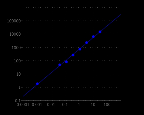
ADP dose response was measured with Amplite® Universal Fluorimetric Kinase Assay Kit in a solid black 384-well plate using a Gemini fluorescence microplate reader (Molecular Devices).
The detection of protein kinase A with the Amplite® Fluorimetric Kinase Assay Kit. The kinase was incubated in the presence of ATP and kemptide peptide substrate for 30 minutes, and ADP generation was detected after 30 minutes incubation using the Amplite® Fluorimetric Kinase Assay Kit.
PLK1-mediated phosphorylation suppressed NDR1 activity to ensure proper spindle orientation. (a)Immunoblot of mitotic HeLa cell lysates expressing GFP-NDR1WT, GFP-NDR13A or GFP-NDR13E, with anti-GFP and anti-pT444-NDR1 antibodies. Quantification is shown on the right. Bars represent means&thinsp;&plusmn;&thinsp;SD, n&thinsp;=&thinsp;3. (b) Kinase activity measure of Flag-NDR1 wild-type or mutants (3A, 3E, K118A and EAIS) purified from asynchronized 293&thinsp;T cells in vitro kinase assay. Full-length blots/gels are presented in&nbsp;Supplementary Fig. S4.(c)and (d) Determination of kinetic parameters of NDR1WT, NDR1K118A and NDR13E. The velocities of kinase assay toward the 10-mer substrate peptide (c) or ATP (d) at varying concentrations were measured by AmpliteTM Universal Fluorimetric Kinase Assay Kit. Data from three independent experiments were analyzed in GraphPad Prism and fitted with Michaelis-Menten equation to extract the kinetic parameters. Bars indicate means&plusmn;SD.(e) Immunoblot analyses of synchronized HeLa cell lysates with (+BI2536) or without (+DMSO) BI2536 treatment, with the antibodies against indicated proteins. &ldquo;M&rdquo; represents mitosis. Quantification is shown on the right. Bars represent means&plusmn;SD from 3 independent experiments. Full-length blots/gels are presented in Supplementary Figure S4.(f) Kinase activity measure of LAP-NDR1 purified from synchronized LAP-NDR1 stable expressing HeLa cells with (+BI2536) or without (+DMSO) BI2536 treatment, or normal HeLa cells (N.C.) in vitro kinase assay. Full-length blots/gels are presented in Supplementary Figure S4.(g) Schematic overview of the experimental procedure of (h). (h) Quantification of spindle angles of mitotic HeLa cells expressing NDR1 siRNA (+siNDR1) or not, with or without BI2536 treatment, as described in (g). Immunofluorescence images were obtained and analyzed as in Fig. 1e. &ldquo;N.C.&rdquo; represents cells with neither transfection nor BI2536 treatment. Bars indicate means&plusmn;SEM from analyses of no less than 40 cells with deformed spindles. Two-tailed student&rsquo;s t-test for p-value calculation.(i) Schematic overview of the experimental procedure of (j).(j) Scatter plots of spindle angles in mitotic HeLa cells transfected with NDR1 siRNA plus various siRNA-resistant NDR1 constructs, as described in (i). Immunofluorescence images were obtained and analyzed as in Figure 1e. Bars indicate means&plusmn;SEM from analyses of 40 cells. Two-tailed student&rsquo;s t-test for p-value calculation. Source: <strong>Regulation of NDR1 activity by PLK1 ensures proper spindle orientation in mitosis </strong>by Yan et al., <em>Scientific Reports</em>, June 2015.
Dimethylation of K191 on PLK1 attenuates its kinase activity. GST-PLK1 was incubated with 6× His-tagged Aurora A plus Bora in the presence of 1 mM SAM for in vitro methylation assay. Kinetics of PLK1 kinase activity in the presence of Aurora A and SET7/9 with increased concentration of ATP. Data represent mean ± SEM from three independent experiments. Source: <b>Methylation of PLK1 by SET7/9 ensures accurate kinetochore–microtubule dynamics</b> by Yu, Ruoying et al., <em>Journal of Molecular Cell Biology</em>, June 2020.
Arrhythmic variant CaM-E140G decreases kinase activity and autophosphorylation of CaMKIIδ. A, quantification of phosphorylation activity of CaMKIIδ using Amplite universal fluorimetric kinase assay kit. (B-left panel), measurement of the relative levels of CaMKIIδ Thr287 autophosphorylation. GST-CaMKIIδ was incubated with CaM variants and ATP for 0 min, 5 min, 15 min, 30 min, and 60 min at room temperature. CaM-WT or CaM-E140G recombinant proteins were used as CaMKIIδ activators. The reaction was terminated using SDS-containing solution, and samples were analyzed by Western blotting and densitometry analysis. (B-right panel), representative blots for CaM-WT and CaM-E140G samples. Phosphorylated proteins (phospho-Thr287 antibody) were normalized to total CaMKIIδ protein (GST antibody). Experiments were performed at least in triplicates. Data are expressed as mean ± s.e.m. Differences between groups were determined using a two-tailed unpaired Student t test. p-values are represented by stars with ∗∗p < 0.01 and ∗∗∗∗p < 0.0001. CaM, calmodulin. Source: <b>Calmodulin variant E140G associated with long QT syndrome impairs CaMKIIδ autophosphorylation and L-type calcium channel inactivation</b>  by Ohm Prakash, Nitika Gupta, Amy Milburn, Liam McCormick, Vishvangi Deugi, Pauline Fisch, Jacob Wyles, N Lowri Thomas, Svetlana Antonyuk, Caroline Dart, and Nordine Helassa. <em>Journal of Biological Chemistry</em>, January 2023.