logo
AAT Bioquest

Amplite® Fluorimetric Catalase Assay Kit *Red Fluorescence*

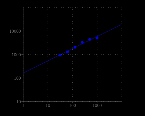
Catalase is a common antioxidant heme-containing redox enzyme found in nearly all living organisms that are exposed to oxygen. The enzyme is concentrated in the peroxisome subcellular organelles. Hydrogen peroxide is an ROS that is a toxic product of normal aerobic metabolism and pathogenic ROS production involving oxidase and superoxide dismutase reactions. By preventing the excessive buildup of H2O2, catalase allows important cellular processes which produce H2O2 as a by-product to take place safely. The Amplite® Fluorimetric Catalase Assay Kit provides a quick and sensitive method for the measurement of catalase activity. It can be performed in a convenient 96-well or 384-well microtiter-plate format and easily adapted to automation without a separation step. Catalase reacts with H2O2 to produce water and oxygen (O2). Amplite® Red also reacts with H2O2 to generate a red fluorescent product. Therefore the reduction in fluorescence intensity is proportional to catalase activity. The Amplite® Red substrate used in the assay enables a dual recordable mode. The fluorescent signal can be easily read by either a fluorescence microplate reader or an absorbance microplate reader. With the Amplite® Fluorimetric Catalase Assay Kit, we have detected as little as 30 mU/mL catalase in a 100 µL reaction volume.
Example protocol

AT A GLANCE

Protocol Summary
  1. Prepare Catalase standards and/or test samples (50 µL)
  2. Add H2O2 Assay Buffer (50 µL)
  3. Incubate at room temperature for 10 - 30 minutes
  4. Add Catalase Assay Mixture (50 µL)
  5. Incubate at room temperature for 10 - 30 minutes
  6. Monitor fluorescence increase at Ex/Em = 540/590 nm (Cutoff = 570nm) 
Important      Thaw all the kit components at room temperature before starting the experiment. The component A is unstable in the presence of thiols such as DTT and β-mercaptoethanol. The final concentration of the thiols higher than 10 µM would significantly decrease the assay dynamic range. NADH and glutathione (reduced form: GSH) may interfere with the assay.

PREPARATION OF STOCK SOLUTIONS

Unless otherwise noted, all unused stock solutions should be divided into single-use aliquots and stored at -20 °C after preparation. Avoid repeated freeze-thaw cycles.

1. Amplite™ Red Substrate stock solution (200X)
Add 65 µL of DMSO (Component F) into the vial of Amplite™ Red (Component A) to make 200X AmpliteTM Substrate stock solution. The stock solution should be used promptly.

2. HRP stock solution (100 U/mL)
Add 200 µL of Assay Buffer (Component C) into the vial of Horseradish Peroxidase (Component D) to make 100 U/mL HRP stock solution.

3. H2O2 stock solution (10 mM)
Add 10 µL of 3% H2O2 (0.88 M, Component B) into 870 µL of Assay Buffer (Component C) to make 10 mM H2O2 stock solution.
Note     The diluted H2O2 stock solution is not stable. The unused portion should be discarded.


4. H2O2 assay buffer (1X)
Add 5 µL of 10 mM H2O2 stock solution into 5 mL of Assay Buffer (Component C) to make 1X H2O2 assay buffer.

5. Catalase standard solution (2 U/mL)
Add 2 µL of 1000 U/mL Catalase Standard (Component E) into 1000 µL of Assay Buffer (Component C) to make 2 U/mL Catalase standard solution.

PREPARATION OF STANDARD SOLUTION

For convenience, use the Serial Dilution Planner:
https://www.aatbio.com/tools/serial-dilution/11306


Catalase standard
Take 2 U/mL Catalase standard solution (CS7) and perform 1:2 serial dilutions to get serially diluted Catalase standard (CS6 - CS1) with Assay Buffer (Component C).

PREPARATION OF WORKING SOLUTION

Add 25 μL of 200X Amplite™ Red substrate stock solution and 15 μL of 100 U/mL HRP stock solution into 5.0 mL of Assay Buffer (Component C) and mix well to prepare Amplite™ Red working solution.
Note     Keep from light.

SAMPLE EXPERIMENTAL PROTOCOL

Table 1.Layout of Catalase standards and test samples in a solid black 96-well microplate. CS= Catalase Standards (CS1 - CS7, 0.031 to 2 U/mL), BL=Blank Control, TS=Test Samples.
BLBLTSTS
CS1CS1......
CS2CS2......
CS3CS3
CS4CS4
CS5CS5
CS6CS6
CS7CS7
Table 2. Reagent composition for each well.
WellVolumeReagents
CS1 - CS750 µLSerial Dilution (0.031 to 2 U/mL)
BL50 µLAssay Buffer (Component C)
TS50 µLtest sample
  1. Prepare Catalase standards (CS), blank controls (BL), and test samples (TS) according to the layout provided in Tables 1 and 2. For a 384-well plate, use 25 µL of reagent per well instead of 50 µL.
  2. Add 50 µL of H2O2 assay buffer to each well of Catalase standard, blank control, and test samples to make the total Catalase assay volume of 100 µL/well. For a 384-well plate, add 25 µL of H2O2 assay buffer into each well instead, for a total volume of 50 µL/well.
  3. Incubate the reaction at room temperature for 15 to 30 minutes, protected from light.
  4. Add 50 µL of Amplite™ Red working solution into each well of Catalase standard, blank control, and test samples to make the total assay volume of 150 µL/well. For a 384-well plate, add 25 µL of Amplite™ Red working solution into each well instead, for a total volume of 75 µL/well.
  5. Incubate the reaction at room temperature for 15 to 30 minutes, protected from light.
  6. Monitor the fluorescence increase with a fluorescence plate reader at Excitation = 540 ± 10, Emission = 590 ± 10 nm (Cutoff = 570 nm) (optimal Ex/Em = 540/590 nm).
    Note     The contents of the plate can also be transferred into a white clear bottom plate and read by an absorbance microplate reader at the wavelength of 576 ± 5 nm. The absorption detection has lower sensitivity compared to fluorescence reading. 

Spectrum

Citations

View all 7 citations: Citation Explorer
Erinacine A-Enriched Hericium erinaceus Mycelium Ethanol Extract Lessens Cellular Damage in Cell and Drosophila Models of Spinocerebellar Ataxia Type 3 by Improvement of Nrf2 Activation
Authors: Wu, Yu-Ling and Sun, Hai-Lun and Chang, Jui-Chih and Lin, Wei-Yong and Chen, Pei-Yin and Chen, Chin-Chu and Lee, Li-Ya and Li, Chien-Chun and Hsieh, Mingli and Chen, Haw-Wen and others,
Journal: Antioxidants (2024): 1495
Stabilizing and Prolonging the Antioxidant Activity of Catalase Using Nondegradable Nanoparticles.
Authors: Murayama, Shuhei and Sasaki, Rikuto and Sakaguchi, Soma and Takaki, Takashi and Kato, Masaru
Journal: Sensors \& Materials (2023)
Glutathione limits RUNX2 oxidation and degradation to regulate bone formation
Authors: Hu, Guoli and Yu, Yilin and Sharma, Deepika and Pruett-Miller, Shondra M and Ren, Yinshi and Zhang, Guo-Fang and Karner, Courtney M
Journal: JCI insight (2023)
A novel approach for the prevention of ionizing radiation-induced bone loss using a designer multifunctional cerium oxide nanozyme
Authors: Wei, Fei and Neal, Craig J and Sakthivel, Tamil Selvan and Fu, Yifei and Omer, Mahmoud and Adhikary, Amitava and Ward, Samuel and Ta, Khoa Minh and Moxon, Samuel and Molinari, Marco and others,
Journal: Bioactive materials (2023): 547--565
In Vitro Efficacy and Molecular Mechanism of Curcumin Analog in Pathological Regulation of Spinocerebellar Ataxia Type 3
Authors: Wu, Yu-Ling and Chang, Jui-Chih and Chao, Yi-Chun and Chan, Hardy and Hsieh, Mingli and Liu, Chin-San
Journal: Antioxidants (2022): 1389

References

View all 94 references: Citation Explorer
Screening for cytochrome p450 reactivity by harnessing catalase as reporter enzyme
Authors: Rabe KS, Spengler M, Erkelenz M, Muller J, G and ubert VJ, Hayen H, Niemeyer CM.
Journal: Chembiochem (2009): 751
New conjugates of superoxide dismutase/catalase mimetics with cyclodestrins
Authors: Lanza V, Vecchio G.
Journal: J Inorg Biochem (2009): 381
Overexpression of catalase delays G0/G1- to S-phase transition during cell cycle progression in mouse aortic endothelial cells
Authors: Onumah OE, Jules GE, Zhao Y, Zhou L, Yang H, Guo Z.
Journal: Free Radic Biol Med (2009): 1658
Novel homo- and hetero-nuclear copper(II) complexes of tetradentate Schiff bases: synthesis, characterization, solvent-extraction and catalase-like activity studies
Authors: Dede B, Karipcin F, Cengiz M.
Journal: J Hazard Mater (2009): 1148
Covalent conjugation of tetrameric bovine liver catalase to liposome membranes for stabilization of the enzyme tertiary and quaternary structures
Authors: Yoshimoto M, Sakamoto H, Shirakami H.
Journal: Colloids Surf B Biointerfaces (2009): 281
Page updated on December 17, 2024

Ordering information

Price
Unit size
Catalog Number11306
Quantity
Add to cart

Additional ordering information

Telephone1-800-990-8053
Fax1-800-609-2943
Emailsales@aatbio.com
InternationalSee distributors
Bulk requestInquire
Custom sizeInquire
Technical SupportContact us
Purchase orderSend to sales@aatbio.com
ShippingStandard overnight for United States, inquire for international
Request quotation

Spectral properties

Excitation (nm)

571

Emission (nm)

584

Storage, safety and handling

H-phraseH303, H313, H333
Hazard symbolXN
Intended useResearch Use Only (RUO)
R-phraseR20, R21, R22
UNSPSC12171501

Platform

Fluorescence microplate reader

Excitation540 nm
Emission590 nm
Cutoff570 nm
Recommended plateSolid black

Components

Catalase dose response was measured with Amplite® Fluorimetric Catalase Assay Kit in a 96-well black solid plate using a Gemini fluorescence microplate reader (Molecular Devices.  As low as 30 mU/mL catalase can be detected with 30 minutes incubation time (n=3).