logo
AAT Bioquest

Amplite® Colorimetric NAD/NADH Ratio Assay Kit

Nicotinamide adenine dinucleotide (NAD+) and nicotinamide adenine dinucleotide phosphate (NADP+) are two important cofactors found in cells. NADH is the reduced form of NAD+, and NAD+ is the oxidized form of NADH. It forms NADP with the addition of a phosphate group to the 2' position of the adenyl nucleotide through an ester linkage. NADP is used in anabolic biological reactions, such as fatty acid and nucleic acid synthesis, which require NADPH as a reducing agent. The traditional NAD/NADH and NADP/NADPH assays are done by monitoring of NADH or NADPH absorption at 340 nm. This method suffers low sensitivity and high interference since the assay is done in the UV range that requires expensive quartz microplate. Our Amplite® NAD/NADH Ratio Assay Kit provides a convenient method for sensitive detection of NAD, NADH and their ratio. The NADH probe is a chromogenic sensor that has its maximum absorbance at ~460 nm upon NADH reduction. The absorbance increase at ~460 nm is directly proportional to the concentration of NADH in the solution. The NADH probe can recognize NADH in an enzyme-free reaction, and the signal can be easily read by an absorbance microplate reader at ~460 nm. The Amplite® Colorimetric NADH Assay Kit provides a sensitive assay to detect as little as 3 µM NADH in a 100 µL assay volume. The assay can be performed in a convenient 96-well or 384-well microtiter-plate format.
Example protocol

AT A GLANCE

Protocol Summary
  1. Prepare 25 µL of NADH standards and/or test samples
  2. Add 25 µL of NAD Extraction Solution
  3. Incubate at 37oC for 15 minutes
  4. Add 25 µL of Neutralization Solution
  5. Add 75 µL of NAD/NADH working solution
  6. Incubate at RT for 15 minutes to 2 hours
  7. Monitor Absorbance at 460 nm
Important Note

It is highly recommended to incubate the cells with Lysis Buffer (Component G) at 37 °C and use the supernatant for the experiment.

Thaw one of each kit component at room temperature before starting the experiment.

CELL PREPARATION

For guidelines on cell sample preparation, please visit https://www.aatbio.com/resources/guides/cell-sample-preparation.html

PREPARATION OF STOCK SOLUTIONS

Unless otherwise noted, all unused stock solutions should be divided into single-use aliquots and stored at -20 °C after preparation. Avoid repeated freeze-thaw cycles

NADH standard solution (1 mM)

Add 200 µL of 1X PBS buffer into the vial of NADH standard (Component C) to make 1 mM (1 nmol/µL) NADH standard solution.

PREPARATION OF STANDARD SOLUTIONS

For convenience, use the Serial Dilution Planner:
https://www.aatbio.com/tools/serial-dilution/15273

NADH standard
Add 50 µL of 1 mM (1 nmol/µL) NADH standard solution into 450 µL 1X PBS buffer (pH 7.4) to generate 100 µM (100 pmols/µL) NADH standard solution. Then take 100 µM NADH standard solution and perform 1:2 serial dilutions in 1X PBS buffer to get serially diluted NADH standards (NS1 - NS7). Note: Diluted NADH standard solution is unstable, and should be used within 4 hours.

PREPARATION OF WORKING SOLUTION

Add 8 mL of NADH Probe Buffer (Component B-II) to the bottle of NAD/NADH Recycling Enzyme Mix (Component A), and mix well.

Add 2 mL NADH Probe (Component B-I) into the bottle of Component A+B-II and mix well to make NAD/NADH working solution.

Note         This NAD/NADH working solution is enough for 125-200 assays. The working solution is not stable, use it promptly and avoid direct exposure to light.

Note         One can try to use ddH2O such as 500 uL to dissolve the Component A, and then mix with B-II and B-I proportionally to make the NAD/NADH working solution for enough use only.

SAMPLE EXPERIMENTAL PROTOCOL

TOTAL NAD+NADH Assay (avail. 400 assays/kit):

Table 1. Layout of NADH standards and test samples in a white/clear bottom 96-well microplate. NS= NADH Standards (NS1 - NS7, 100 to 1.56 µM), BL=Blank Control, TS=Test Samples.

BLBLTSTS
NS1NS1......
NS2NS2......
NS3NS3  
NS4NS4  
NS5NS5  
NS6NS6  
NS7NS7  

Table 2. Reagent composition for each well. High concentration of NADH (e.g., >100 µM, final concentration) will cause saturated signal and make the calibration curve non-linear.

WellVolumeReagent
NS1 - NS750 µLSerial Dilutions (100 to 1.56 µM)
BL50 µL1X PBS
TS50 µLTest Sample
  1. Prepare NADH standards (NS), blank controls (BL), and test samples (TS) according to the layout provided in Tables 1 and 2.

    Note         Prepare cells or tissue samples as desired. Lysis Buffer (Component G) can be used for lysing the cells for convenience and incubate the cells with Lysis Buffer at 37oC for 15 minutes and use the supernatant for the experiment.
  2. Add 50 µL of NAD/NADH working solution into each well of NADH standard, blank control, and test samples to make the total NAD/NADH assay volume of 100 µL/well.
  3. Incubate the reaction at room temperature for 15 minutes to 2 hours, protected from light.
  4. Monitor the absorbance increase with an absorbance plate reader at 460 nm.
NAD/NADH RATIO Assay (avail. 250 assays/kit):

Table 3. Layout of NADH standards and test samples in a white/clear 96-well microplate. NS= NADH Standards (NS1 - NS7, 100 to 1.56 µM); BL=Blank Control; TS=Test Samples; TS (NAD) = Test Samples treated with NAD Extraction Solution (Component D) for 10 to 15 minutes, then neutralized by Neutralization Solution (Component E).

BLBLTSTSTS (NAD)TS (NAD)
NS1NS1......  
NS2NS2......  
NS3NS3    
NS4NS4    
NS5NS5    
NS6NS6    
NS7NS7    

Table 4. Reagent compositions for each well. High concentration of NADH (e.g., >100 µM, final concentration) will cause saturated signal and make the calibration curve non-linear.

NADH Standard Blank Control

Test Sample (NAD+NADH)

Test Sample (NAD Extract)
Serial Dilutions: 25 µL 1X PBS: 25 µL Test Sample: 25 µL Test Sample: 25 µL
Component F: 25 µL Component F: 25 µL Component F: 25 µL Component D: 25 µL
Incubate at 37 °C for 10 to 15 minutes Incubate at 37 °C for 10 to 15 minutes Incubate at 37 °C for 10 to 15 minutes Incubate at 37 °C for 10 to 15 minutes
Component F: 25 µL Component F: 25 µL Component F: 25 µL Component E: 25 µL
Total: 75 µL Total: 75 µL Total: 75 µL Total: 75 µL
  1. Refer to Tables 3 & 4 for compositions of each well.

    1. For NAD Extraction (NAD amount): Add 25 µL of NAD Extraction Solution (Component D) into the wells of NAD/NADH containing test samples. Incubate at 37oC for 10 to 15 minutes, then add 25 µL of Neutralization Solution (Component E) to neutralize the NAD extracts as described in Tables 3 & 4.
    2. For Total NAD and NADH (Total amount): Add 25 µL of Extraction Control Solution (Component F) into the wells of NADH standards and NAD/NADH containing test samples. Incubate at room 37oC for 10 to 15 minutes, and then add 25 µL of Extraction Control Solution (Component F) as described in Tables 3 and 4.

      Note         Prepare cells or tissue samples as desired. Lysis Buffer (Component G) can be used for lysing the cells for convenience.
  2. Add 75 µL of NAD/NADH working solution into each well of NADH standard, blank control, and test samples (NAD/NADH), and test sample (NAD Extract) to make the total assay volume of 150 µL/well.

  3. Incubate the reaction at room temperature for 15 minutes to 2 hours (We tested 60 minutes in the figure shown), protected from light

  4. Monitor the absorbance increase with an absorbance plate reader at 460 nm.

Citations

View all 81 citations: Citation Explorer
Regulatory Roles of NAMPT and NAD+ Metabolism in Uterine Leiomyoma Progression: Implications for ECM Accumulation, Stemness, and Microenvironment
Authors: Chiang, Yi-Fen and Huang, Ko-Chieh and Huang, Tsui-Chin and Chen, Hsin-Yuan and Ali, Mohamed and Al-Hendy, Ayman and Huang, Pei-Shen and Hsia, Shih-Min
Journal: Redox Biology (2024): 103411
Neuroglobin protects dopaminergic neurons in a Parkinson’s cell model by interacting with mitochondrial complex NDUFA10
Authors: Liang, Xiaomei and Wen, Yutong and Feng, Cuilian and Xu, Lan and Xian, Ying and Xie, Haiting and Huang, Jianou and Huang, Yihong and Zhao, Xiaodong and Gao, Xiaoya
Journal: Neuroscience (2024)
Deregulation of mitochondrial reverse electron transport alters the metabolism of reactive oxygen species and NAD+/NADH and presents a therapeutic target in Alzheimer’s disease
Authors: Rimal, Suman and Li, Wen and Khaket, Tejinder Pal and Li, Yu and Tantray, Ishaq and Li, Yanping and Bhurtel, Sunil and Grinberg, Lea T and Spina, Salvatore and Sillero, Maria Inmaculada Cobos and others,
Journal: (2024)
Exposure to fluoride exacerbates the cognitive deficit of diabetic patients living in areas with endemic fluorosis, as well as of rats with type 2 diabetes induced by streptozotocin via a mechanism that may involve excessive activation of the poly (ADP rib
Authors: Xiang, Jie and Qi, Xiao-Lan and Cao, Kun and Ran, Long-Yan and Zeng, Xiao-Xiao and Xiao, Xiao and Liao, Wei and He, Wen-Wen and Hong, Wei and He, Yan and others,
Journal: Science of The Total Environment (2023): 169512
The Exposure to 2.45 GHz Electromagnetic Radiation Induced Different Cell Responses in Neuron-like Cells and Peripheral Blood Mononuclear Cells
Authors: Bertuccio, Maria Paola and Acri, Giuseppe and Ientile, Riccardo and Caccamo, Daniela and Curr{\`o}, Monica
Journal: Biomedicines (2023): 3129
Page updated on November 20, 2024

Ordering information

Price
Unit size
Catalog Number15273
Quantity
Add to cart

Additional ordering information

Telephone1-800-990-8053
Fax1-800-609-2943
Emailsales@aatbio.com
InternationalSee distributors
Bulk requestInquire
Custom sizeInquire
Technical SupportContact us
Purchase orderSend to sales@aatbio.com
ShippingStandard overnight for United States, inquire for international
Request quotation

Storage, safety and handling

Certificate of OriginDownload PDF
H-phraseH303, H313, H333
Hazard symbolXN
Intended useResearch Use Only (RUO)
R-phraseR20, R21, R22
UNSPSC12352200

Platform

Absorbance microplate reader

Absorbance460 nm
Recommended plateClear bottom

Components

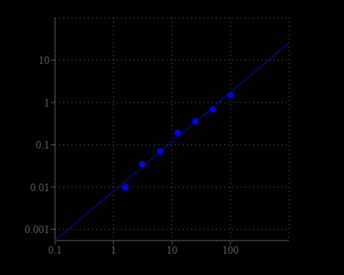
Amplite® Colorimetric NAD/NADH Ratio Assay Kit is used to measure total NAD/NADH amount and in a white/clear 96-well microplate using a SpectraMax microplate reader (Molecular devices).
Amplite® Colorimetric NAD/NADH Ratio Assay Kit is used to measure total NAD/NADH amount and NAD/NADH ratio in a white/clear 96-well microplate using a SpectraMax microplate reader (Molecular devices). Right: Total NADH and NAD dose response; Left: NAD/NADH ratio: Equal amount of NAD and NADH mixture was treated with or without NAD extraction solution for 15 minutes, and then neutralized with extraction solution at room temperature. The signal was read at 460 nm. NAD/NADH molar ratio is calculated based on the absorbance shown in the figure 1 (left).