Acridine Orange
Acridine orange is a cell permeable, nucleic acid dye that exhibits green fluorescence for dsDNA and red fluorescence for RNA or ssDNA.
Acridine orange is a derivative of acridine that was originally developed as a way to quickly label and identify bacteria. Over time, it has become established as a fluorophore for staining nucleic acids. Chemically, it has a neutral charge and is weakly basic, which facilitates its diffusion across cell membranes. As such, it can be used to stain live cells. Once in the cytoplasm, it tends to accumulate and be retained by acidic cellular compartments with low pH. Because of this, acridine orange has found use as a stain for lysosomes.
Nucleic acid staining
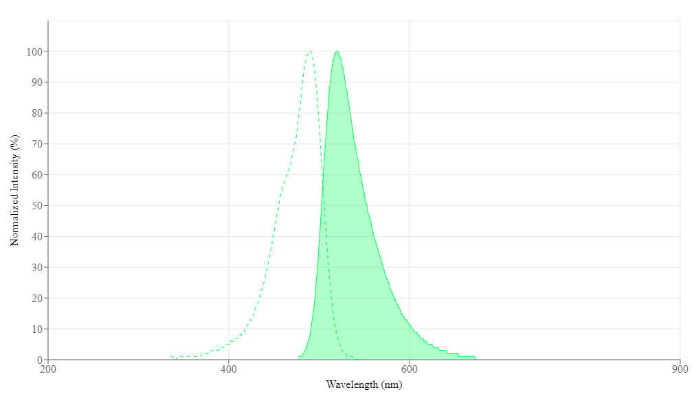
Given its weakly basic nature, acridine orange also binds to dsDNA, ssDNA and RNA, which are acidic. The binding to dsDNA is thought to occur by way of intercalation. This results in a low concentration of acridine orange around the nucleic acid, yielding a green fluorescence with an excitation peak at 490 nm and an emission peak at 520 nm. On the other hand, binding of acridine orange to RNA is thought to occur through electrostatic forces. This results in a higher concentration of the fluorophore and a red fluorescence, with an excitation peak at 460 nm and an emission peak at 640 nm.Acridine orange and propidium iodide
Acridine orange is sometimes used in conjugation with propidium iodide to assess cell viability. Since acridine orange is cell permeable, it will stain live and dead cells. Propidium iodide, on the other hand, is membrane impermeant and will only stain dead cells. Used together, live and dead cells can be quantified.Nonyl acridine orange
Nonyl acridine orange is a derivative of acridine orange that specifically accumulates in, and is used to study, mitochondria.Further reading
- Eriksson, I., Vainikka, L., Persson, H. L., & Öllinger, K. (2023). Real-time monitoring of lysosomal membrane permeabilization using acridine orange. Methods and Protocols, 6(4), 72.
- McMaster, G. K., & Carmichael, G. G. (1977). Analysis of single-and double-stranded nucleic acids on polyacrylamide and agarose gels by using glyoxal and acridine orange. Proceedings of the National Academy of Sciences, 74(11), 4835-4838.
- Plemel, J. R., Caprariello, A. V., Keough, M. B., Henry, T. J., Tsutsui, S., Chu, T. H., ... & Stys, P. K. (2017). Unique spectral signatures of the nucleic acid dye acridine orange can distinguish cell death by apoptosis and necroptosis. Journal of Cell Biology, 216(4), 1163-1181.
- Robbins, E., & Marcus, P. I. (1963). Dynamics of acridine orange-cell interaction: I. Interrelationships of acridine orange particles and cytoplasmic reddening. The Journal of cell biology, 18(2), 237-250.
- Wolf, M. K., & Aronson, S. B. (1961). Growth, fluorescence and metachromasy of cells cultured in the presence of acridine orange. Journal of Histochemistry & Cytochemistry, 9(1), 22-29.
Calculators
Common stock solution preparation
Table 1. Volume of Water needed to reconstitute specific mass of Acridine orange to given concentration. Note that volume is only for preparing stock solution. Refer to sample experimental protocol for appropriate experimental/physiological buffers.
0.1 mg | 0.5 mg | 1 mg | 5 mg | 10 mg | |
1 mM | 331.323 µL | 1.657 mL | 3.313 mL | 16.566 mL | 33.132 mL |
5 mM | 66.265 µL | 331.323 µL | 662.647 µL | 3.313 mL | 6.626 mL |
10 mM | 33.132 µL | 165.662 µL | 331.323 µL | 1.657 mL | 3.313 mL |
Molarity calculator
Enter any two values (mass, volume, concentration) to calculate the third.
Mass (Calculate) | Molecular weight | Volume (Calculate) | Concentration (Calculate) | Moles | ||||
/ | = | x | = |
Spectrum
Open in Advanced Spectrum Viewer
Product family
Name | Excitation (nm) | Emission (nm) |
Thiazole orange *CAS 107091-89-4* | 514 | 533 |
Thiazole orange *10 mM in DMSO* | 514 | 533 |
CytoTell™ Orange | 541 | 560 |
LysoBrite™ Orange | 543 | 565 |
References
View all 56 references: Citation Explorer
Spectral studies of N-nonyl acridine orange in anionic, cationic and neutral surfactants
Authors: Wiosetek-Reske AM, Wysocki S.
Journal: Spectrochim Acta A Mol Biomol Spectrosc (2006): 1118
Authors: Wiosetek-Reske AM, Wysocki S.
Journal: Spectrochim Acta A Mol Biomol Spectrosc (2006): 1118
Re: The evaluation of micronucleus frequency by acridine orange fluorescent staining in peripheral blood of rats treated with lead acetate. (Mutagenesis, 20, 411-415, 2005)
Authors: Celik A, Ogenler O, Comelekoglu U.
Journal: Mutagenesis (2006): 267
Authors: Celik A, Ogenler O, Comelekoglu U.
Journal: Mutagenesis (2006): 267
Improvement of the acridine orange-protein-surfactant system for protein estimation based on aromatic ring stacking effect of sodium dodecyl benzene sulphonate
Authors: Wang F, Yang J, Wu X, Wang X, Guo C, Jia Z.
Journal: Luminescence (2006): 186
Authors: Wang F, Yang J, Wu X, Wang X, Guo C, Jia Z.
Journal: Luminescence (2006): 186
Vesicle disruption, plasma membrane bleb formation, and acute cell death caused by illumination with blue light in acridine orange-loaded malignant melanoma cells
Authors: Hiruma H, Katakura T, Takenami T, Igawa S, Kanoh M, Fujimura T, Kawakami T.
Journal: J Photochem Photobiol B. (2006)
Authors: Hiruma H, Katakura T, Takenami T, Igawa S, Kanoh M, Fujimura T, Kawakami T.
Journal: J Photochem Photobiol B. (2006)
The use of acridine orange base (AOB) as molecular probe to characterize nonaqueous AOT reverse micelles
Authors: Falcone RD, Correa NM, Biasutti MA, Silber JJ.
Journal: J Colloid Interface Sci (2006): 356
Authors: Falcone RD, Correa NM, Biasutti MA, Silber JJ.
Journal: J Colloid Interface Sci (2006): 356
Page updated on November 21, 2024