logo
AAT Bioquest

PhosphoWorks™ Fluorimetric Pyrophosphate Assay Kit *Blue Fluorescence*

Pyrophosphate (PPi) is produced by a number of biochemical reactions, such as ATP hydrolysis, DNA and RNA polymerizations, cyclic AMP formation by the enzyme adenylate cyclase and the enzymatic activation of fatty acids to form their coenzyme A esters. Our PhosphoWroks™ Pyrophosphate Assay Kit provides the most robust spectrophotometric method for measuring pyrophosphate. This kit uses our proprietary fluorogenic pyrophosphate sensor that has its fluorescence intensity proportionally dependent upon the concentration of pyrophosphate. Our assay is much easier and more robust than the enzyme-coupling pyrophosphate methods that require at least two enzymes for their pyrophosphate detections. The kit provides all the essential components for assaying pyrophosphate. This kit has been successfully used in high throughput screening (HTS). Please inquire special HTS bulk package discount for the screening of >10,000 assays.
Example protocol

AT A GLANCE

Protocol Summary
  1. Prepare Pyrophosphate standards and/or test samples (50 µL)
  2. Add Pyrophosphate working solution (50 µL)
  3. Incubate at room temperature for 10 to 30 minutes
  4. Monitor fluorescence intensity at Ex/Em = 316/456 nm (Cutoff = 420 nm)
Important Note

Thaw all the four components at room temperature before use.

PREPARATION OF STOCK SOLUTIONS

Unless otherwise noted, all unused stock solutions should be divided into single-use aliquots and stored at -20 °C after preparation. Avoid repeated freeze-thaw cycles

PPi Sensor stock solution (200X)

Add 50 µL of DMSO (Component D) into the vial of PPi Sensor (Component B) to make 200X PPi Sensor stock solution. Protect from light.
Note        25 µL of the PPi Sensor Stock Solution is enough for one 96-well plate.

Pyrophosphate standard solution (1 mM)

Add 10 µL of 50 mM Pyrophosphate Standard (Component C) into 490 µL of ddH2O or 50 mM Hepes buffer (pH 7) to make 1 mM Pyrophosphate standard solution.

PREPARATION OF STANDARD SOLUTIONS

For convenience, use the Serial Dilution Planner:
https://www.aatbio.com/tools/serial-dilution/21611

Pyrophosphate standard
Add 50 μL of 1 mM Pyrophosphate standard solution into 450 μL of ddH2O or 50 mM Hepes buffer to get 100 μM Pyrophosphate standard solution (PS7). Take 100 μM Pyrophosphate standard solution and perform 1:3 serial dilutions in ddH2O or 50 mM Hepes buffer to get serially diluted Pyrophosphate standards (PS6 - PS1).

PREPARATION OF WORKING SOLUTION

Add 25 μL of 200X PPi Sensor stock solution to 5 mL of Assay Buffer (Component A) and mix well to make PPi working solution.
Note        Due to the high sensitivity of this assay to PPi, it is important to use PPi-free labware and reagents. DTT ≥ 1 mM will increase the background, MgCl2 ≥ 2 mM will decrease the response.

SAMPLE EXPERIMENTAL PROTOCOL

Table 1. Layout of Pyrophosphate standards and test samples in a solid black 96-well microplate. PS = Pyrophosphate Standard (PS1 - PS7, 0.14 to 100 µM), BL = Blank Control, TS = Test Sample.

BLBLTSTS
PS1PS1......
PS2PS2......
PS3PS3
PS4PS4
PS5PS5
PS6PS6
PS7PS7

Table 2. Reagent composition for each well.

WellVolumeReagent
PS1 - PS750 µL

Serial Dilutions (0.14 to 100 µM)

BL50 µLAssay Buffer
TS50 µLtest sample
  1. Prepare Pyrophosphate standards (PS), blank controls (BL), and test samples (TS) according to the layout provided in Tables 1 and 2. For a 384-well plate, use 25 µL of reagent per well instead of 50 µL.
  2. Add 50 µL of PPi working solution to each well of Pyrophosphate standard, blank control, and test samples to make the total Pyrophosphate assay volume of 100 µL/well. For a 384-well plate, add 25 µL of PPi working solution into each well instead, for a total volume of 50 µL/well. Mix the reagents thoroughly.
  3. Incubate at room temperature for 10 to 30 minutes.
  4. Monitor the fluorescence increase with a fluorescence plate reader at Ex/Em = 316/456 nm (Cutoff = 420 nm).

Citations

View all 12 citations: Citation Explorer
Inorganic Pyrophosphate at Serum Concentration May Not Be Able to Inhibit Mineralization: A Study in Aqueous Solutions and Serum
Authors: Cheng, Yuxuan and Ru, Jing and Feng, Chaobo and Liu, Xiaohao and Zeng, Hua and Tan, Shuo and Chen, Xi and Chen, Feng and Lu, Bing-Qiang
Journal: ACS Omega (2024)
AaTAS1 and AaMFS1 Genes for Biosynthesis or Efflux Transport of Tenuazonic Acid and Pathogenicity of Alternaria alternata
Authors: Sun, Fan and Cao, Xueqiang and Yu, Dianzhen and Hu, Dongqiang and Yan, Zheng and Fan, Yingying and Wang, Cheng and Wu, Aibo
Journal: Molecular Plant-Microbe Interactions (2022): 416--427
Augmented fibroblast growth factor-23 secretion in bone locally contributes to impaired bone mineralization in chronic kidney disease in mice
Authors: Andrukhova, Olena and Sch{\"u}ler, Christiane and Bergow, Claudia and Petric, Alexandra and Erben, Reinhold G
Journal: Frontiers in endocrinology (2018): 311
RANKL Expression Is Increased in Circulating Mononuclear Cells of Patients with Calcific Aortic Stenosis
Authors: Rattazzi, Marcello and Faggin, Elisabetta and Bertacco, Elisa and Buso, Roberta and Puato, Massimo and Plebani, Mario and Zaninotto, Martina and Condotta, Davide and Zoppellaro, Giacomo and Pagliani, Leopoldo and others, undefined
Journal: Journal of Cardiovascular Translational Research (2018): 1--10
Biological studies and target engagement of the 2-C-methyl-d-erythritol 4-phosphate cytidylyltransferase (IspD)-targeting antimalarial agent (1 R, 3 S)-MMV008138 and analogs
Authors: Ghavami, Maryam and Merino, Emilio F and Yao, Zhong-Ke and Elahi, Rubayet and Simpson, Morgan E and Fern{\'a}ndez-Murga, Maria L and Butler, Joshua H and Casasanta, Michael A and Krai, Priscilla M and Totrov, Maxim M and others,
Journal: ACS infectious diseases (2017): 549--559

References

View all 136 references: Citation Explorer
Cell-surface-localized ATP detection with immobilized firefly luciferase
Authors: Nakamura M, Mie M, Funabashi H, Yamamoto K, Ando J, Kobatake E.
Journal: Anal Biochem (2006): 61
Ca2+ oscillations stimulate an ATP increase during fertilization of mouse eggs
Authors: Campbell K, Swann K.
Journal: Dev Biol (2006): 225
An efficient method for quantitative determination of cellular ATP synthetic activity
Authors: Hara KY, Mori H.
Journal: J Biomol Screen (2006): 310
Basic evaluation for new antimicrobial susceptibility testing of Mycobacterium leprae by bioluminescence assay (ATP method)
Authors: Yamazaki T, Gidoh M, Matsuoka M.
Journal: Nihon Hansenbyo Gakkai Zasshi (2006): 227
ATP bioluminescence assay for estimation of microbial populations of fresh-cut melon
Authors: Ukuku DO, Sapers GM, Fett WF.
Journal: J Food Prot (2005): 2427
Page updated on November 23, 2024

Ordering information

Price
Unit size
Catalog Number21611
Quantity
Add to cart

Additional ordering information

Telephone1-800-990-8053
Fax1-800-609-2943
Emailsales@aatbio.com
InternationalSee distributors
Bulk requestInquire
Custom sizeInquire
Technical SupportContact us
Purchase orderSend to sales@aatbio.com
ShippingStandard overnight for United States, inquire for international
Request quotation

Storage, safety and handling

H-phraseH303, H313, H333
Hazard symbolXN
Intended useResearch Use Only (RUO)
R-phraseR20, R21, R22
UNSPSC12352200

Platform

Fluorescence microplate reader

Excitation316 nm
Emission456 nm
Cutoff420 nm
Recommended plateSolid black

Components

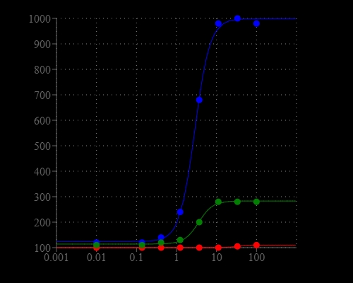
Pyrophosphate, ATP and phosphate dose responses were measured with PhosphoWorks™ Fluoremetric Pyrophosphate Assay Kit in a solid black 96-well plate using a fluorescence microplate reader.