logo
AAT Bioquest

Cell Meter™ Nuclear Apoptosis Assay Kit *Green Fluorescence Optimized for Flow Cytometry*

Our Cell Meter™ assay kits are a set of tools for monitoring cell viability. There are a variety of parameters that can be used for monitoring cell viability. This particular kit is designed to monitor cell apoptosis through measuring the apoptotic chromatin condensation. The compacted chromatin of apoptotic cells binds higher amounts of nuclear dye compared to the healthy cells. Our Cell Meter™ Nuclear Apoptosis Assay Kit provides all the essential components with an optimized assay method for the detection of apoptosis in cells with condensed nuclei. This fluorometric assay is based on the detection of the DNA contents in cells using our proprietary non-fluorescent dye that becomes strongly fluorescent upon binding to DNA. In normal cells, Nuclear Green™ is not cell permeable, however, in apoptotic cells, Cells with compromised plasma membranes or with impaired/no cell metabolism are unable to prevent the dye from entering the cell. Once inside the cell, the dyes bind to intracellular DNA producing highly fluorescent complexes which identify the cells as non-viable cells. The staining with Nuclear Green; DCS1 can be measured using a flow cytometer (FL1 channel) or fluorescence microscope (FITC filter set). The kit can be used with our other apoptosis reagents, such as Our Cell Meter™ NIR Mitochondria Membrane Potential Detection Kit (Cat# 22802), for multi-parametric study of cell viability and apoptosis. The kit is optimized for screening of apoptosis activators and inhibitors.
Example protocol

AT A GLANCE

Protocol summary

  1. Prepare cells with test compounds at a density of 5 × 10⁵ to 1 × 10⁶ cells/mL
  2. Add 5 µL of 200X Nuclear Green™ DCS1 into 1 mL of cell solution
  3. Incubate the cells in a 37°C, 5% CO2 incubator for 30 to 60 minutes
  4. Pellet the cells and resuspend the cells in 1 mL of growth medium
  5. Analyze the fluorescence intensity using flow cytometer with FL1 channel

Important notes
Thaw all the kit components at room temperature before starting the experiment.

SAMPLE EXPERIMENTAL PROTOCOL

  1. For each sample, prepare cells in 1 mL of warm medium or buffer of your choice at a density of 5 × 10⁵ to 1 × 10⁶ cells/mL. Note: Each cell line should be evaluated on an individual basis to determine the optimal cell density for apoptosis induction.

  2. Treat cells with test compounds for a desired period of time to induce apoptosis.

  3. Add 5 µL of 200X Nuclear Green™ DCS1 (Component A) into the treated cells.

  4. Incubate the cell solution in a 37°C, 5% CO2 incubator for 30 to 60 minutes. Note: For adherent cells, gently lift the cells with 0.5 mM EDTA to keep the cells intack, and wash the cells once with serum-containing media prior to the incubation with Nuclear Green™ DCS1 dye-loading solution. The appropriate incubation time depends on the individual cell type and cell concentration used. Optimize the incubation time for each experiment.

  5. Optional: Centrifuge the cells at 1000 rpm for 4 minutes, and then re-suspend cells in 1 mL of Assay Buffer (Component B) or buffer of your choice.

  6. Monitor the fluorescence intensity using a flow cytometer with FL1 channel (Ex/Em = 490/525 nm). Gate on the cells of interest, excluding debris.

Spectrum

References

View all 93 references: Citation Explorer
Curcumin induces cell cycle arrest and apoptosis in human osteosarcoma (HOS) cells
Authors: Lee DS, Lee MK, Kim JH.
Journal: Anticancer Res (2009): 5039
Real-time in vivo imaging of retinal cell apoptosis after laser exposure
Authors: Schmitz-Valckenberg S, Guo L, Maass A, Cheung W, Vugler A, Moss SE, Munro PM, Fitzke FW, Cordeiro MF.
Journal: Invest Ophthalmol Vis Sci (2008): 2773
Morphological study on apoptosis Hela cells induced by soyasaponins
Authors: Xiao JX, Huang GQ, Zhu CP, Ren DD, Zhang SH.
Journal: Toxicol In Vitro (2007): 820
Effects of neuraminidase on apoptosis of blood lymphocytes in rats with implanted Morris tumor
Authors: Grata-Borkowska U, Steciwko A, Pokorski M, Drobnik J, Gasiorowski K, Pirogowicz I, Cieslar-Marczak E.
Journal: J Physiol Pharmacol (2007): 253
Effects of Chinese herbal recipe Weichang'an in inducing apoptosis and related gene expression in human gastric cancer grafted onto nude mice
Authors: Zhao AG, Yang JK, You SF, Li T, Zhao HL, Gu Y, Tang LD, Qiu JX.
Journal: Zhong Xi Yi Jie He Xue Bao (2007): 287
Page updated on January 10, 2025

Ordering information

Price
Unit size
Catalog Number22811
Quantity
Add to cart

Additional ordering information

Telephone1-800-990-8053
Fax1-800-609-2943
Emailsales@aatbio.com
InternationalSee distributors
Bulk requestInquire
Custom sizeInquire
Technical SupportContact us
Purchase orderSend to sales@aatbio.com
ShippingStandard overnight for United States, inquire for international
Request quotation

Spectral properties

Excitation (nm)

503

Emission (nm)

527

Storage, safety and handling

H-phraseH303, H313, H333
Hazard symbolXN
Intended useResearch Use Only (RUO)
R-phraseR20, R21, R22
UNSPSC12352200

Platform

Flow cytometer

Excitation488 nm laser
Emission530, 30 nm filter
Instrument specification(s)FITC channel

Components

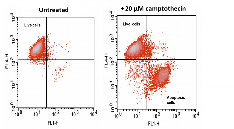
The increase in fluorescence intensity of Nuclear Green™ DCS1 with the addition of Camptothecin in Jurkat cells. Jurkat cells were treated overnight without (Left) or with 20 μM camptothecin (Right) in a 37 oC, 5% CO2 incubator, and then dye loaded with Nuclear Green™ DCS1 for 60 minutes. At the end of 15 minutes of Nuclear Green™ DCS1 dye loading, MitoLite™ NIR (Cat. # 22802) was added for multicolor analysis. The fluorescence intensity of Nuclear Green™ DCS1 and MitoLite™ NIR was measured with a FACSCalibur (Becton Dickinson, San Jose, CA) flow cytometer using FL1 channel (Nuclear Green™ DCS1) and FL4 channel (MitoLite™ NIR).