logo
AAT Bioquest

Amplite® Universal Fluorimetric MMP Activity Assay Kit *Red Fluorescence*

The matrix metalloproteinases (MMPs) constitute a family of zinc-dependent endopeptidases that function within the extracellular matrix. These enzymes are responsible for the breakdown of connective tissues and are important in bone remodeling, the menstrual cycle and repair of tissue damage. While the exact contribution of MMPs to certain pathological processes is difficult to assess, MMPs appear to play a key role in the development of arthritis as well as in the invasion and metastasis of cancer. MMPs tend to have multiple substrates, with most family members having the ability to degrade different types of collagen along with elastin, gelatin and fibronectin. It is quite difficult to find a substrate that is selective to a single MMP enzyme. This kit is designed to check the general activity of a MMP enzyme. It can also be used to screening MMP inhibitors when a purified MMP enzyme is used. We also offer a few MMP enzyme of high activity.
Example protocol

AT A GLANCE

Protocol summary

  1. Prepare appropriate controls or test samples (50 µL)
  2. Pre-incubate for 10 -15 minutes 
  3. Add MMP RedTM Substrate working solution (50 µL)
  4. Skip incubation for kinetic reading or incubate 30 minutes - 1 hour for end point reading
  5. Monitor fluorescence intensity at Ex/Em = 540/590 nm (Cutoff = 570 nm)

Important notes
Thaw all the kit Components at room temperature before starting the experiment. 

PREPARATION OF WORKING SOLUTION

1. APMA working solution (2mM, 2X):
Dilute 1 M APMA (Component B) with Assay Buffer (Component C) at 1:500 to make 2 mM, 2X APMA working solution . Note: APMA belongs to organic mercury. Handle with care! Dispose it according to local regulations.

2. MMP RedTM Substrate working solution
:
Add 50 µL of MMP RedTM Substrate (Component A) to 5 mL of Assay Buffer (Component C) and mix well to make MMP RedTM Substrate working solution. Note: MMP RedTM Substrate working solution is enough for one 96-well plate (100 assays).

3. MMP dilution:
Dilulte MMPs to an appropriate concentration with Assay Buffer (Component C) if purified MMP is used. Note: Pro-MMP needs to be activated before use.  Avoid vigorously vortexing the enzyme.

4. Inhibitors and compounds dilutions:
Make dilutions of known MMPs inhibitors and test compounds as desired if you are screening MMPs inhibitors.

For guidelines on cell sample preparation, please visit
https://www.aatbio.com/resources/guides/cell-sample-preparation.html

SAMPLE EXPERIMENTAL PROTOCOL

Table 1. Protocols for pro-MMP activation

MMPsActivated by Treating with
MMP-1 (collagenase)1 mM APMA (diluted component C) at 37 °C for 3 hr.
MMP-2 (gelatinase)1 mM APMA (diluted component C) at 37 °C for 1 hr.
MMP-3 (stromelysin)1 mM APMA (diluted component C) at 37 °C for 24 hr.
MMP-7 (matrilysin, PUMP-1)1 mM APMA (diluted component C) at 37 °C for 20 min-1 hr.
MMP-8 (neutrophil collagenase)1 mM APMA (diluted component C) at 37 °C for 1 hr.
MMP-9 (92 kDa gelatinase)1 mM APMA (diluted component C) at 37 °C for 2 hr.
MMP-10 (stromelysin 2)1 mM APMA (diluted component C) at 37 °C for 24 hr.
MMP-11 (stromelysin-3)Already in active form. No APMA treatment is necessary.
MMP-12 (macrophage elastase)1 mM APMA (diluted component C) at 37 °C for 2 hr.
MMP-13 (collagenase-3)1 mM APMA (diluted component C) at 37 °C for 40 min.
MMP-141 mM APMA (diluted component C) at 37 °C for 2-3 hr.


Table 2.
 Layout of the samples in a solid black 96-well microplate.  SC=Substrate Control, IC=Inhibitor Control, VC=Vehicle Control, TC=Test Compound Control, TS=Test Samples. 

SCSC......
ICIC......
VCVC  
TCTC  
TSTS  
......  


Table 3. Reagent composition for each well. Some strongly fluorescent test compounds may result in false-positive results.

WellVolumeReagent
SC50 µLAssay Buffer 
IC

50 µL

MMP dilution and known MMPs inhibitor

VC50 µL

MMP dilution and vehicle used to deliver test compound

TC50 µLAssay Buffer and test compound
TS50 µL

MMP dilution with test compound

  1. Prepare MMPs containing biological samples as desired.

  2. Incubate the MMP containing-samples or purified MMPs with equal volume of 2 mM APMA working solution (2X). Refer to Table 1 for incubation time. Activate MMP immediately before the experiment. Note: Keep enzyme-containing samples on ice. Avoid vigorously vortexing the enzyme. Prolonged storage of the activated enzyme will deactivate the enzyme. Note: For enzyme activation, it is preferably activated at higher protein concentration. After activation, you may further dilute the enzyme.

  3. Prepare Subtrate Control (SC), Inhibitor Control (IC), Vehicle Control (VC), Test Compound Control (TC) and Test Samples (TS) according to the layout provided in Table 2 and Table 3.

  4. Pre-incubate the plate at a desired temperature for the enzyme reaction (e.g. 25 °C or 37 °C) for 10-15 min if you are screening MMPs inhibitors.

  5. Add 50 µL/well (96-well plate) or 20 µL/well (384-well plate) of MMP Red™ Substrate working solution to the sample and control wells of the assay plate. Mix the reagents well.

  6. Monitor the fluorescence intensity with a fluorescence plate reader at Ex/Em = 540/590 nm (Cutoff = 570 nm).

    For kinetic reading: 
    Immediately start measuring fluorescence intensity continuously and record data every 5 minutes for 30 minutes.

    For end-point reading: Incubate the reaction at a desired temperature for 30 to 60 minutes, protected from light.  Mix the reagents well, and then measure the fluorescence intensity.

Spectrum

Product family

Citations

View all 16 citations: Citation Explorer
MiRNA29a-3p Negatively Regulates ISL1--Integrin $\beta$1 Axis to Suppress Gastric Cancer Progression
Authors: She, Ziwei and Dong, Haosheng and Li, Yang and Chen, Ping and Zhou, Chunyan and Wang, Weiping and Jia, Zhuqing and Shi, Qiong
Journal: Experimental Cell Research (2024): 114288
Targeting cancer-derived extracellular vesicles by combining CD147 inhibition with tissue factor pathway inhibitor for the management of urothelial cancer cells
Authors: Boddu, Vijay Kumar and Zamzow, Piet and Kramer, Mario Wolfgang and Merseburger, Axel S and Gorantla, Sivahari Prasad and Klinger, Matthias and Cramer, Lena and Sauer, Thorben and Gemoll, Timo and von Bubnoff, Nikolas and others,
Journal: Cell Communication and Signaling (2024): 1--14
RNA-binding protein-regulated fibronectin is essential for EGFR-activated metastasis of head and neck squamous cell carcinoma
Authors: Shieh, Jiunn-Min and Chang, Ting-Wei and Wang, Jing-He and Liang, Song-Ping and Kao, Pei-Lu and Chen, Liang-Yi and Yen, Chia-Jui and Chen, Yun-Ju and Chang, Wen-Chang and Chen, Ben-Kuen
Journal: The FASEB Journal (2023): e23206
Influence of Dinitrosyl Iron Complexes on the Activity of Enzymes, Indicators of Cardiovascular Diseases
Authors: Akentieva, Natalia and Sanina, Natalia and Gizatullin, Artur and Shkondina, Natalia and Andreeva, Anna and Shram, Stanislav and Aldoshin, Sergei
Journal: (2022)
Zinc ions regulate opening of tight junction favouring efflux of macromolecules via the GSK3β/snail-mediated pathway
Authors: Xiao, Ruyue and Yuan, Lan and He, Weijiang and Yang, Xiaoda
Journal: Metallomics (2018)
Page updated on November 21, 2024

Ordering information

Price
Unit size
Catalog Number13511
Quantity
Add to cart

Additional ordering information

Telephone1-800-990-8053
Fax1-800-609-2943
Emailsales@aatbio.com
InternationalSee distributors
Bulk requestInquire
Custom sizeInquire
Technical SupportContact us
Purchase orderSend to sales@aatbio.com
ShippingStandard overnight for United States, inquire for international
Request quotation

Spectral properties

Excitation (nm)

545

Emission (nm)

572

Storage, safety and handling

H-phraseH303, H313, H333
Hazard symbolXN
Intended useResearch Use Only (RUO)
R-phraseR20, R21, R22
UNSPSC12352200

Platform

Fluorescence microplate reader

Excitation540 nm
Emission590 nm
Cutoff570 nm
Recommended plateSolid black

Components

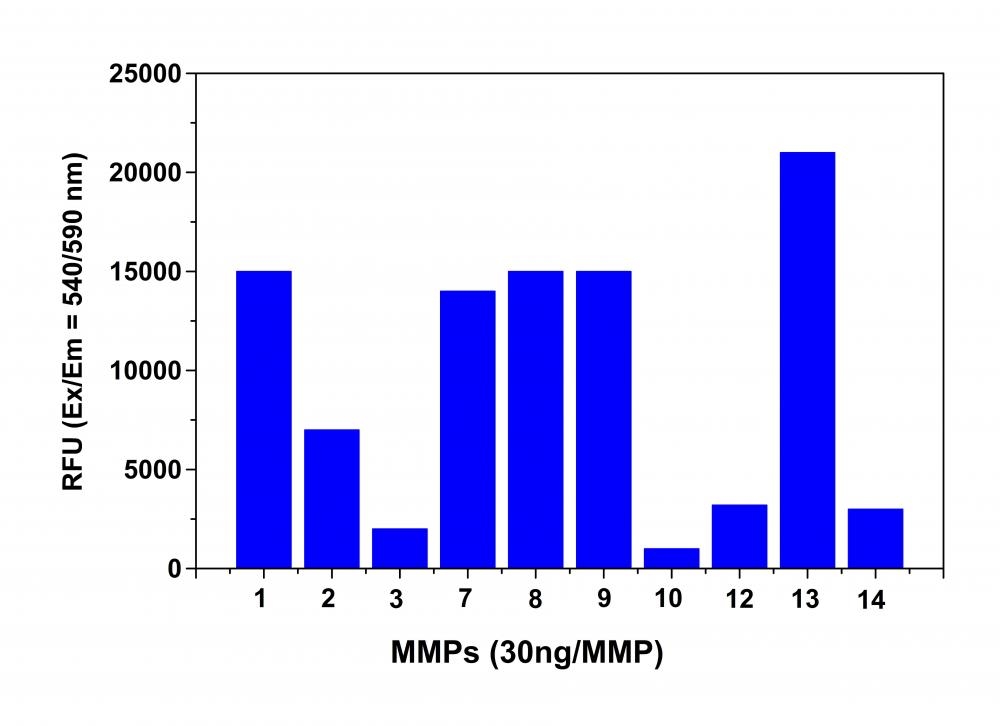
Detection of MMPs activity using Amplite® Universal Fluorimetric MMP Activity Assay Kit. The fluorescence signal was monitored one hour after the start of the reaction. The reading from all wells was subtracted with the reading from substrate control, which contains MMP Red™ substrate but no MMPs. The MMP Red™ substrate can detect the activity of sub-nanogram of all MMPs (n=3).