Amplite® Fluorimetric Xanthine Assay Kit
Example protocol
AT A GLANCE
Protocol summary
- Prepare xanthine standards or test samples (50 µL)
- Add xanthine working solution (50 µL)
- Incubate at room temperature for 30 - 60 min
- Read fluorescence intensity at Ex/Em = 540/590 nm
Important notes
Thaw all the kit components at room temperature before starting the experiment.
PREPARATION OF STOCK SOLUTION
1. Amplite™ Red substrate stock solution (250X):
Add 40 µL of DMSO (Component F) into the vial of Amplite™ Red substrate (Component A). Note: Amplite™ Red substrate is unstable in the presence of thiols such as dithiothreitol (DTT) and 2-mercaptoethanol. The final concentration of DTT or 2-mercaptoethanol in the reaction should be no higher than 10 µM. The assay should be performed at pH 7 - 8 (pH 7.4 is recommended) as Amplite™ Red is unstable at pH > 8.5.
2. HRP stock solution (500X):
Add 100 µL of Assay Buffer (Component B) into the vial of Horseradish Peroxidase (Component C).
3. Xanthine oxidase (XO) stock solution (100X):
Add 100 µL of Assay Buffer (Component B) into the vial of Xanthine Oxidase (Component E) to make Xanthine Oxidase (XO) stock solution (100X).
PREPARATION OF STANDARD SOLUTION
For convenience, use the Serial Dilution Planner: https://www.aatbio.com/tools/serial-dilution/13843
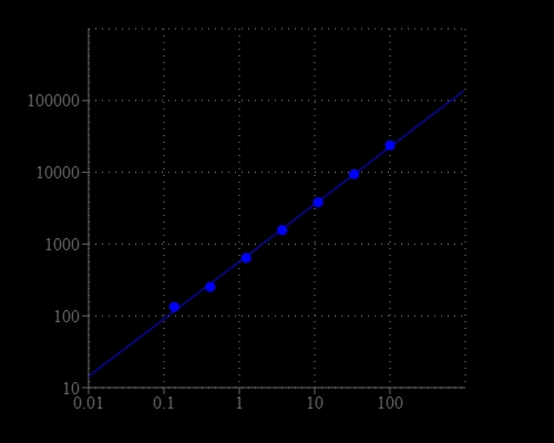
Add 5 µL of Xanthine Standard (Component D) into 995 µL of Assay Buffer (Component B) to get 100 µM xanthine standard solution. Take 200 µL of 100 µM xanthine standard solution to perform 1:3 serial dilutions to get serially diluted xanthine standards (X1 - X7).
PREPARATION OF WORKING SOLUTION
Add 20 μL of Amplite™ Red Substrate stock solution (250X), 10 μL of HRP stock solution (500X), and 50 μL of Xanthine Oxidase stock solution (100X) into 5 mL of Assay Buffer (Component B) to make a total volume of 5.08 mL. Note: Avoid direct exposure to light and use promptly.
SAMPLE EXPERIMENTAL PROTOCOL
Table 1. Layout of Xanthine standards and test samples in a black wall/solid bottom 96-well microplate. X = xanthine standard (X1 - X7, 0.137 to 100 µM); BL = blank control; TS = test sample.
BL | BL | TS | TS |
X1 | X1 | ... | ... |
X2 | X2 | ... | ... |
X3 | X3 | ||
X4 | X4 | ||
X5 | X5 | ||
X6 | X6 | ||
X7 | X7 |
Table 2. Reagent composition for each well.
Well | Volume | Reagent |
X1 - X7 | 50 µL | Serial Dilution (0.137 to 100 µM) |
BL | 50 µL | Assay Buffer (Component B) |
TS | 50 µL | Test Sample |
- Prepare xanthine standards (X), blank controls (BL), and test samples (TS) according to the layout provided in Table 1 and Table 2. For a 384-well plate, use 25 µL of reagent per well instead of 50 µL.
- Add 50 µL of xanthine working solution into each well of the xanthine standards, blank control, and test samples to make the total xanthine assay volume of 100 µL/well. For a 384-well plate, add 25 µL of working solution into each well instead, for a total volume of 50 µL/well.
- Incubate the reaction for 30 to 60 minutes at room temperature, protected from light.
- Monitor the fluorescence increase with with a fluorescence plate reader at Excitation = 530 - 570 nm (optimal at 540 nm), Emission = 590 - 600 nm (optimal at 590 nm), cutoff = 570 nm.
References
Authors: Martin DE, De Almeida JF, Henry MA, Khaing ZZ, Schmidt CE, Teixeira FB, Diogenes A.
Journal: J Endod (2014): 51
Authors: Kitamura K, Maruyama K, Hamano S, Kishi T, Kawakami T, Takahashi Y, Onodera S.
Journal: J Toxicol Sci (2014): 71
Authors: Yin B, Deng J, Peng X, Long Q, Zhao J, Lu Q, Chen Q, Li H, Tang H, Zhang Y, Yao S.
Journal: Analyst (2013): 6551
Authors: Thorn RM, Robinson GM, Reynolds DM.
Journal: Antimicrob Agents Chemother (2013): 2216
Authors: Setlow B, Yu J, Li YQ, Setlow P.
Journal: Lett Appl Microbiol (2013): 259