logo
AAT Bioquest

Amplite® Fluorimetric NAD/NADH Ratio Assay Kit *Red Fluorescence*

Nicotinamide adenine dinucleotide (NAD+) and nicotinamide adenine dinucleotide phosphate (NADP+) are two important cofactors found in cells. NADH is the reduced form of NAD+, and NAD+ is the oxidized form of NADH. It forms NADP with the addition of a phosphate group to the 2' position of the adenyl nucleotide through an ester linkage. NADP is used in anabolic biological reactions, such as fatty acid and nucleic acid synthesis, which require NADPH as a reducing agent. The traditional NAD/NADH and NADP/NADPH assays are done by monitoring of NADH or NADPH absorption at 340 nm. This method suffers low sensitivity and high interference since the assay is done in the UV range that requires expensive quartz microplate. Our Amplite® NAD/NADH Ratio Assay Kit provides a convenient method for sensitive detection of NAD, NADH and their ratio. The enzymes in the system specifically recognize NAD/NADH in an enzyme cycling reaction. There is no need to purify NAD/NADH from sample mix. The enzyme cycling reaction significantly increases detection sensitivity. In addition, this assay has very low background since it is run in the red visible range that significantly reduces the interference from biological samples. The assay has demonstrated high sensitivity and low interference. This Amplite® Fluorimetric NAD/NADH Assay Kit can be performed in a convenient 96-well or 384-well microtiter-plate format and easily adapted to automation with no separation steps required.

Example protocol

AT A GLANCE

Protocol Summary
  1. Prepare 25 µL of NADH standards and/or test samples
  2. Add 25 µL of NADH or NAD Extraction Solution
  3. Incubate at 37oC for 15 minutes
  4. Add 25 µL of NAD or NADH Extraction Solution
  5. Add 75 µL of NAD/NADH working solution
  6. Incubate at RT for 15 minutes to 2 hours
  7. Monitor fluorescence intensity at Ex/Em = 540/590 nm
Important Note

It is highly recommended to incubate the cells with Lysis Buffer (Component G) at 37oC and use the supernatent for the experiment.

Thaw one of each kit component at room temperature before starting the experiment.

CELL PREPARATION

For guidelines on cell sample preparation, please visit https://www.aatbio.com/resources/guides/cell-sample-preparation.html

PREPARATION OF STOCK SOLUTIONS

Unless otherwise noted, all unused stock solutions should be divided into single-use aliquots and stored at -20 °C after preparation. Avoid repeated freeze-thaw cycles

NADH standard solution (1 mM)

Add 200 µL of PBS buffer into the vial of NADH standard (Component C) to have 1 mM (1 nmol/µL) NADH stock solution.

PREPARATION OF STANDARD SOLUTIONS

For convenience, use the Serial Dilution Planner:
https://www.aatbio.com/tools/serial-dilution/15263

NADH standard
Use NADH standard solution and PBS buffer (pH 7.4) to generate 30 µM (30 pmols/µL) NADH standard solution (NS7). Then take the 30 µM NADH standard solution to perform 1:3 serial dilutions to get remaining serially diltued NADH standards (NS6-NS1). Note: Diluted NADH standard solution is unstable, and should be used within 4 hours.

PREPARATION OF WORKING SOLUTION

Add 10 mL of NADH Sensor Buffer (Component B) to the bottle of NAD/NADH Recycling Enzyme Mixture (Component A), and mix well.
Note         This NAD/NADH working solution is enough for 125 assays in 96-well plate. The working solution is not stable, use it promptly and avoid direct exposure to light.

SAMPLE EXPERIMENTAL PROTOCOL

Table 1. Layout of NADH standards and test samples in a solid black 96-well microplate. NS = NADH standard (NS1-NS7, 0.03 to 30 µM); BL = blank control; TS = test sample; TS(NADH) = test sample treated with NADH Extraction Solution, then neutralized by NAD Extraction Solution ; TS(NAD) = test sample treated with NAD Extraction Solution , then neutralized by NADH Extraction Solution.

BLBLTSTSTS(NADH)TS(NADH)TS(NAD)TS(NAD)
NS1NS1..................
NS2NS2..................
NS3NS3
NS4NS4
NS5NS5
NS6NS6
NS7NS7

Table 2. Reagent composition for each well. Take note that high concentration of NADH (e.g., >300 µM, final concentration) may cause reduced fluorescence signal due to the over oxidation of NADH sensor (to a non-fluorescent product).

WellReagentTreatmentTreatmentTotal

NS1-NS7

Serial Dilution (0.03 to 30 µM) (25 µL)

Component F (25 µL)

Incubate at 37 °C for 10 to 15 minutes

Component F (25 µL)

75 µL

BL

PBS (25 µL)

Component F (25 µL)

Incubate at 37 °C for 10 to 15 minutes

Component F (25 µL)

75 µL

TS (NAD+NADH)

Sample (25 µL)

Component F (25 µL)

Incubate at 37 °C for 10 to 15 minutes

Component F (25 µL)

75 µL

TS (NADH extract)

Sample (25 µL)

Component D (25 µL)

Incubate at 37 °C for 10 to 15 minutes

Component E (25 µL)

75 µL

TS (NAD extract)

Sample (25 µL)

Component E (25 µL)

Incubate at 37 °C for 10 to 15 minutes

Component D (25 µL)

75 µL

Prepare NADH standards (NS), blank controls (BL), test samples (TS), test samples treated with NADH Extraction Solution (TS(NADH)), and test samples treated with NAD Extraction Solution (TS(NAD)) according to the layout described in Tables 1 and 2.
Note         Prepare cells or tissue samples as desired.
Note         Incubate the cells with Lysis Buffer for 15 mins at 37oC and use the supernatant for the experiment.

For NAD Extraction (NAD): Add 25 µL of NAD Extraction Solution (Component E) into the wells of NAD/NADH containing test samples. Incubate at 37oC for 10 to 15 minutes, then add 25 µL of NADH Extraction Solution (Component D) to neutralize the NAD extracts as described in Tables 1 & 2.

For Total NAD and NADH: Add 25 µL of NAD/NADH Control Solution (Component F) into the wells of NADH standards and NAD/NADH containing test samples. Incubate at 37oC for 10 to 15 minutes, and then add 25 µL of Control Solution (Component F) as described in Tables 1 and 2.

For NADH Extraction (NADH): Add 25 µL of NADH Extraction Solution (Component D) into the wells of NAD/NADH containing test samples. Incubate at 37oC for 10 to 15 minutes, then add 25 µL of NAD Extraction Solution (Component E) to neutralize the NADH extracts as described in Tables 1 & 2. Note: In healthy mammalian cells, there is more NAD comparing to NADH, so one can simply use total NAD and NADH minus the NAD to calculate the amount of NADH.

  1. Add 75 µL of NAD/NADH working solution into each well of NADH standard, blank control, and test samples to make the total NADH assay volume of 150 µL/well.

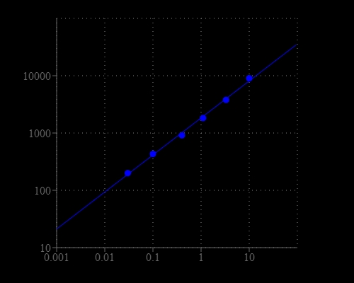
  2. Incubate the reaction at room temperature for 15 minutes to 2 hours (We tested 60 minutes in the figure shown), protected from light.
  3. Monitor the fluorescence increase with a fluorescence plate reader at Ex/Em = 540/590 nm (cutoff 570 nm).

    Note         The contents of the plate can also be transferred to a white clear bottom plate and read by an absorbance microplate reader at the wavelength of 576 ± 5 nm. However, the absorption detection has lower sensitivity compared to fluorescence reading.

NAD/NADH assay calculator

The standard curve needed for detection of NAD, NADH and their ratio is generated by measuring the absorbances of a known NADH standard. This standard curve can then be used to calculate the concentration of any unknown sample.

This tool will generate a linear regression model for any given experimental data set with which to detect NAD/NADH concentration for the NAD/NADH Assay.
Begin Calculation

Citations

View all 84 citations: Citation Explorer
Breast cancer cells utilize T3 to trigger proliferation through cellular Ca2+ modulation
Authors: Tawfik, Ines and Schlick, Katharina and Ostaku, Julian and Bresilla, Doruntina and Gabrijel{\v{c}}i{\v{c}}, Sonja and Gottschalk, Benjamin and Sokolowski, Alwin and Malle, Ernst and Kalinova, Katarina and Hirtl, Martin and others,
Journal: Cell Communication and Signaling (2024): 533
The coupling of mitoproteolysis and oxidative phosphorylation enables tracking of an active mitochondrial state through MitoTimer fluorescence
Authors: Xie, Yinyin and Zhang, Yannan and Sun, Aina and Peng, Yamei and Hou, Weikang and Xiang, Cong and Zhang, Guoxin and Lai, Beibei and Hou, Xiaoshuang and Zheng, Fangfang and others,
Journal: Redox biology (2022): 102447
Elucidating the mechanism by which synthetic helper peptides sensitize Pseudomonas aeruginosa to multiple antibiotics
Authors: Xia, Yushan and Cebri{\'a}n, Rub{\'e}n and Xu, Congjuan and Jong, Anne de and Wu, Weihui and Kuipers, Oscar P
Journal: PLoS pathogens (2021): e1009909
Rapamycin Modulates the Proinflammatory Memory-Like Response of Microglia Induced by BAFF
Authors: Wang, Jianing and Yang, Chunshu and Hou, Xiaoyu and Xu, Jingyi and Yun, Yang and Qin, Ling and Yang, Pingting
Journal: Frontiers in Immunology (2021): 1649
Page updated on November 21, 2024

Ordering information

Price
Unit size
Catalog Number15263
Quantity
Add to cart

Additional ordering information

Telephone1-800-990-8053
Fax1-800-609-2943
Emailsales@aatbio.com
InternationalSee distributors
Bulk requestInquire
Custom sizeInquire
Technical SupportContact us
Purchase orderSend to sales@aatbio.com
ShippingStandard overnight for United States, inquire for international
Request quotation

Storage, safety and handling

H-phraseH303, H313, H333
Hazard symbolXN
Intended useResearch Use Only (RUO)
R-phraseR20, R21, R22
UNSPSC12352200

Platform

Fluorescence microplate reader

Excitation540 nm
Emission590 nm
Cutoff570 nm
Recommended plateSolid black

Components

NADH response was measured with Amplite® Fluorimetric NAD/NADH Ratio Assay Kit in a 96-well solid black plate using a Gemini microplate reader (Molecular Devices).