logo
AAT Bioquest

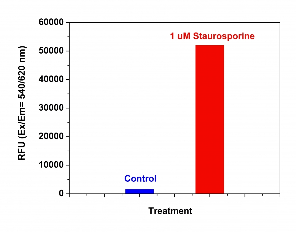
Amplite® Fluorimetric Caspase 3/7 Assay Kit *Red Fluorescence*

Caspases play important roles in apoptosis and cell signaling. The activation of caspase-3 (CPP32/apopain) is important for the initiation of apoptosis. Caspase 3 is also identified as a drug-screening target. Caspase 3 has substrate selectivity for the peptide sequence Asp-Glu-Val-Asp (DEVD). This Amplite® Caspase-3 Assay Kit uses Z-DEVD-ProRed™ as the fluorogenic indicator for assaying caspase-3 activity. Cleavage of R110 peptides by caspases generates strongly red fluorescent ProRed™ that can be monitored fluorimetrically at ~620 nm with excitation of ~530 nm. Z-DEVD-ProRed™ is recognized as the most sensitive red fluorogenic caspase 3/7 substrate. This kit can be used to continuously measure the activities of caspase-3 in cell extracts and purified enzyme preparations using a fluorescence microplate reader or fluorometer. It can also be used with flow cytometry for analyzing cell apoptosis and the activities of caspases 3 and 7.

Example protocol

AT A GLANCE

Protocol summary

  1. Prepare cells with test compounds
  2. Add equal volume of caspase 3/7 working solution
  3. Incubate at room temperature for 1 hour
  4. Monitor fluorescence intensity at Ex/Em = 535/620 nm

Important notes
Thaw Component A, B, C (if desired, Component D) at room temperature before use.

PREPARATION OF STOCK SOLUTION

Unless otherwise noted, all unused stock solutions should be divided into single-use aliquots and stored at -20 °C after preparation. Avoid repeated freeze-thaw cycles.

1. Z-DEVD-ProRed™ stock solution (200X):
Add 65 µL of DMSO (not provided) into the vial of Component A.

2. (Optional) Caspase 3/7 Inhibitor Ac-DEVD-CHO stock solution (1 mM):
Add 100 µL of DMSO directly to the vial of Ac-DEVD-CHO (Component D). This inhibitor can be used to confirm the correlation between fluorescence signal intensity and caspase 3/7-like protease activities.

PREPARATION OF WORKING SOLUTION

Add 50 μL of 200X Z-DEVD-ProRed™ stock solution and 100 μL of 1M DTT solution (Component C) into 10 mL Assay Buffer (Component B) and mix well.


Note: 50 μL of the 200X Z-DEVD-ProRed™ stock solution is enough for 100 assays using a reaction volume of 100 μL per assay.

For guidelines on cell sample preparation, please visit
https://www.aatbio.com/resources/guides/cell-sample-preparation.html

SAMPLE EXPERIMENTAL PROTOCOL

  1. Treat cells by adding 10 µL of 10X test compounds (96-well plate) or 5 µL of 5X test compounds (384-plate) into PBS or desired buffer. For blank wells (medium without the cells), add the same amount of compound buffer.

  2. Incubate the cell plates in an incubator for a desired period of time (3 - 5 hours for Jurkat cells treated with staurosporine) to induce apoptosis.

  3. Add 100 µL/well (96-well plate) or 25 µL/well (384-well plate) of caspase 3/7 working solution.

  4. Incubate the plate at room temperature for at least 1 hour, kept from light. Note: If desired, add 1 µL of the 1 mM stock solution of the caspase 3/7 Inhibitor Ac-DEVD-CHO into selected samples 10 minutes before adding the caspase 3/7 assay working solution at room temperature to confirm the caspase 3/7-like activities.

  5. Monitor the fluorescence intensity at Ex/Em = 535/620 nm (cut off at 610 nm) with either top or bottom read mode. Note: Sometimes, bottom read gives better signal to background ratio, centrifuge cell plate (especially for the nonadherent cells) at 800 rpm for 2 minutes (brake off) if using bottom read mode.

Spectrum

Product family

Citations

View all 18 citations: Citation Explorer
BRPF1 inhibition reduces migration and invasion of metastatic ovarian cancer cells, representing a potential therapeutic target
Authors: Alexandrova, Elena and Smal, Marharyta and Di Rosa, Domenico and Brancaccio, Rosario Nicola and Parisi, Roberto and Russo, Fabio and Tarallo, Roberta and Nassa, Giovanni and Giurato, Giorgio and Weisz, Alessandro and others,
Journal: Scientific Reports (2025): 7602
An engineered Palivizumab IgG2 subclass for synthetic gp130 and fas mediated signaling
Authors: Wittich, Christoph and Ettich, Julia and Hertell, Marcel and Roy, Biswadeep G and Xu, Haifeng C and Floss, Doreen M and Lang, Philipp A and Scheller, J{\"u}rgen
Journal: Journal of Biological Chemistry (2025): 108205
Goji Berry Juice Prevents Tumor Necrosis Factor Alpha-Induced Xerostomia in Human Salivary Gland Cells
Authors: Takakura, Masatoshi and Mizutani, Ayano and Kudo, Mizuki and Ishikawa, Airi and Okamoto, Takuya and Fu, Tong Xuan and Kurimoto, Shin-ichiro and Koike, Yuka and Mishima, Kenji and Tanaka, Junichi and others,
Journal: Biological and Pharmaceutical Bulletin (2024): 138--144
Respiratory syncytial virus--approved mAb Palivizumab as ligand for anti-idiotype nanobody-based synthetic cytokine receptors
Authors: Ettich, Julia and Wittich, Christoph and Moll, Jens M and Behnke, Kristina and Floss, Doreen M and Reiners, Jens and Christmann, Andreas and Lang, Philipp A and Smits, Sander HJ and Kolmar, Harald and others,
Journal: The Journal of Biological Chemistry (2023)
Respiratory syncytial virus (RSV)-approved monoclonal antibody Palivizumab as ligand for anti-idiotype nanobody-based synthetic cytokine receptors
Authors: Ettich, Julia and Wittich, Christoph and Moll, Jens M and Behnke, Kristina and Floss, Doreen M and Reiners, Jens and Christmann, Andreas and Lang, Philipp A and Smits, Sander HJ and Kolmar, Harald and others,
Journal: Journal of Biological Chemistry (2023): 105270

References

View all 67 references: Citation Explorer
Quantitative measurement of caspase-3 activity in a living starfish egg
Authors: Sakaue M, Motoyama Y, Yamamoto K, Shiba T, Teshima T, Chiba K.
Journal: Biochem Biophys Res Commun (2006): 878
Serofendic acid, a neuroprotective substance derived from fetal calf serum, inhibits mitochondrial membrane depolarization and caspase-3 activation
Authors: Kume T, Taguchi R, Katsuki H, Akao M, Sugimoto H, Kaneko S, Akaike A.
Journal: Eur J Pharmacol (2006): 69
Multiparameter measurement of caspase 3 activation and apoptotic cell death in NT2 neuronal precursor cells using high-content analysis
Authors: Fennell M, Chan H, Wood A.
Journal: J Biomol Screen (2006): 296
Asymmetric dimethylarginine induces apoptosis via p38 MAPK/caspase-3-dependent signaling pathway in endothelial cells
Authors: Jiang DJ, Jia SJ, Dai Z, Li YJ.
Journal: J Mol Cell Cardiol (2006): 529
Diallyl Trisulfide Induces Apoptosis of Human Gastric Cancer Cell Line MGC803 Through Caspase-3 Pathway.
Authors: Xiao XL, Peng J, Su Q, Xiang SL, Tang GH, Huang YS, Zhou XT.
Journal: Ai Zheng (2006): 1247
Page updated on March 11, 2025

Ordering information

Price
Unit size
Catalog Number13504
Quantity
Add to cart

Additional ordering information

Telephone1-800-990-8053
Fax1-800-609-2943
Emailsales@aatbio.com
InternationalSee distributors
Bulk requestInquire
Custom sizeInquire
Technical SupportContact us
Purchase orderSend to sales@aatbio.com
ShippingStandard overnight for United States, inquire for international
Request quotation

Spectral properties

Excitation (nm)

532

Emission (nm)

619

Storage, safety and handling

H-phraseH303, H313, H333
Hazard symbolXN
Intended useResearch Use Only (RUO)
R-phraseR20, R21, R22
UNSPSC12352200

Platform

Fluorescence microplate reader

Excitation535 nm
Emission620 nm
Cutoff610 nm
Recommended plateSolid black

Components

Detection of Caspase 3/7 Activities in Jurkat cells. Jurkat cells were seeded on the same day at 200,000 cells/90 uL/well in a Costar black wall/clear bottom 96-well plate. The cells were treated with staurosporine at the final concentration of 1 uM for 5 hours while the untreated cells were used as control. The caspase 3/7 assay solution (100 uL/well) was added and incubated at room temperature for 1 hour. The fluorescence intensity was measured at Ex/Em = 540/620 nm with FlexStation fluorescence microplate reader (Molecular Devices).