logo
AAT Bioquest

Amplite® Colorimetric Acetylcholinesterase Assay Kit

Acetylcholinesterase, also known as AChE, is an enzyme that degrades (through its hydrolytic activity) the neurotransmitter acetylcholine, producing choline and an acetate group. It is mainly found at neuromuscular junctions and cholinergic synapses in the central nervous system, where its activity serves to terminate synaptic transmission. AChE has a very high catalytic activity- each molecule of AChE degrades about 5000 molecules of acetylcholine per second. Acetylcholinesterase is also found on the red blood cell membranes, where it constitutes the Yt blood group antigen. Acetylcholinesterase exists in multiple molecular forms, which possess similar catalytic properties, but differ in their oligomeric assembly and mode of attachment to the cell surface. This Amplite® Colorimetric Acetylcholinesterase Assay Kit provides a convenient method for the detecting AChE activity. The kit uses DTNB to quantify the thiolcholine produced from the hydrolysis of acetylthiolcholine by AChE. The absoprtion intensity of DTNB adduct is proportional to the formation of thiolcholine, thus the AChE activity.
Example protocol

AT A GLANCE

Protocol summary

  1. Prepare AChE working solution (50 µL)
  2. Add AChE standards or AChE test samples (50 µL)
  3. Incubate at room temperature for 10 - 30 minutes
  4. Monitor absorbance at 410 ± 5 nm

Important notes
Thaw all the kit components at room temperature before starting the experiment.

PREPARATION OF STOCK SOLUTION

Unless otherwise noted, all unused stock solutions should be divided into single-use aliquots and stored at -20 °C after preparation. Avoid repeated freeze-thaw cycles.

1. DTNB stock solution (20X):
Add 0.6 mL of Assay Buffer (Component B) into the vial of DTNB (Component A) to make 20X DTNB stock solution. Keep from light. Note: DNTB is not easy to dissolve, it is normal to see the cloudiness of the solution. One can use either the supernatant or the mixture for the experiment.

2. Acetylthiocholine stock solution (20X):
Add 0.6 mL of ddH2O into the vial of Acetylthiocholine (Component C) to make 20X Acetylthiocholine stock solution. 

3. Acetylcholinesterase standard solution (50 U/mL):
Add 100 µL of ddH2O with 0.1% BSA into the vial of Acetylcholinesterase Standard (Component D) to make 50 U/mL Acetylcholinesterase standard solution.

PREPARATION OF STANDARD SOLUTION

Acetylcholinesterase standard

For convenience, use the Serial Dilution Planner: https://www.aatbio.com/tools/serial-dilution/11400

Add 20 µL of 50 U/mL Acetylcholinesterase standard solution to 980 µL of Assay Buffer (Component B) to generate 1000 mU/mL Acetylcholinesterase standard solution (AS7). Then take 1000 mU/mL Acetylcholinesterase standard solution (AS7) and perform 1:3 serial dilutions in Assay Buffer (Component B) to get serially diluted Acetylcholinesterase standards (AS6 - AS1). Note: Diluted acetylcholinesterase standard solution is unstable and should be used within 4 hours.

PREPARATION OF WORKING SOLUTION

Add 250 μL of 20X DTNB stock solution and 250 μL of 20X Acetylthiocholine stock solution into 4.5 mL of Assay Buffer (Component B) to make a total volume of 5 mL AChE working solution. Keep from light.

For guidelines on cell sample preparation, please visit
https://www.aatbio.com/resources/guides/cell-sample-preparation.html

SAMPLE EXPERIMENTAL PROTOCOL

Table 1. Layout of Acetylcholinesterase standards and test samples in a white/clear bottom 96-well microplate. AS=Acetylcholinesterase Standards (AS1 - AS7, 1 to 1000 mU/mL); BL=Blank Control; TS=Test Samples.

BLBLTSTS
AS1AS1......
AS2AS2......
AS3AS3  
AS4AS4  
AS5AS5  
AS6AS6  
AS7AS7  

Table 2. Reagent composition for each well.

WellVolumeReagent
AS1 - AS750 µLSerial Dilutions (1 to 1000 mU/mL)
BL50 µLAssay Buffer (Component B)
TS50 µLtest sample
  1. Prepare Acetylcholinesterase standards (AS), blank controls (BL), and test samples (TS) according to the layout provided in Tables 1 and 2. For a 384-well plate, use 25 µL of reagent per well instead of 50 µL. Note: Treat cells or tissue samples as desired.

  2. Add 50 µL of AChE working solution to each well of Acetylcholinesterase standard, blank control, and test samples to make the total Acetylcholinesterase assay volume of 100 µL/well. For a 384-well plate, add 25 µL of AChE working solution into each well instead, for a total volume of 50 µL/well.

  3. Incubate the reaction for 10 to 30 minutes at room temperature, protected from light.

  4. Monitor the absorbance increase with an absorbance microplate reader at 410 ± 5 nm.

Citations

View all 60 citations: Citation Explorer
Isolation of new indole alkaloid triglucoside from the aqueous extract of Uncaria rhynchophylla
Authors: Koseki, Yuta and Nishimura, Hiroaki and Asano, Ryuji and Aoki, Katsuyuki and Shiyu, Li and Sugiyama, Ryosuke and Yamazaki, Mami
Journal: Journal of Natural Medicines (2024): 1--8
Identification of Potent and Selective Acetylcholinesterase/Butyrylcholinesterase Inhibitors by Virtual Screening
Authors: Xu, Tuan and Li, Shuaizhang and Li, Andrew J and Zhao, Jinghua and Sakamuru, Srilatha and Huang, Wenwei and Xia, Menghang and Huang, Ruili
Journal: Journal of Chemical Information and Modeling (2023)
Cell shape characteristics of human skeletal muscle cells as a predictor of myogenic competency: A new paradigm towards precision cell therapy
Authors: Desprez, Charlotte and Danovi, Davide and Knowles, Charles H and Day, Richard M
Journal: Journal of Tissue Engineering (2023): 20417314221139794
Amelioration of AlCl3-induced Memory Loss in the Rats by an Aqueous Extract of Guduchi, a Medhya Rasayana
Authors: Jamadagni, Shrirang B and Ghadge, Pooja M and Tambe, Mukul S and Srinivasan, Marimuthu and Prasad, Goli Penchala and Jamadagni, Pallavi S and Prasad, Shyam Baboo and Pawar, Sharad D and Gurav, Arun M and Gaidhani, Sudesh N and others,
Journal: Pharmacognosy Magazine (2023): 09731296221145063
Synergistic protective effect of Beta vulgaris with meso-2, 3-dimercaptosuccinic acid against lead-induced neurotoxicity in male rats
Authors: Shaban, Nadia Z and Abd El-Kader, Sara E and Mogahed, Fayed AK and El-Kersh, Mohamed AL and Habashy, Noha H
Journal: Scientific Reports (2021): 1--18

References

View all 41 references: Citation Explorer
Activation of the nitric oxide-cGMP pathway reduces phasic contractions in neonatal rat bladder strips via protein kinase G
Authors: Artim DE, Kullmann FA, Daugherty SL, Wu HY, de Groat WC.
Journal: Am J Physiol Renal Physiol (2009): F333
Protective effect of stilbenes containing extract-fraction from Cajanus cajan L. on Abeta(25-35)-induced cognitive deficits in mice
Authors: Ruan CJ, Si JY, Zhang L, Chen DH, Du GH, Su L.
Journal: Neurosci Lett (2009): 159
Platelet activating factor (PAF) and activity of its biosynthetic and catabolic enzymes in blood and leukocytes of male patients with newly diagnosed heart failure
Authors: Detopoulou P, Nomikos T, Fragopoulou E, Antonopoulou S, Kotroyiannis I, Vassiliadou C, Panagiotakos DB, Chrysohoou C, Pitsavos C, Stefanadis C.
Journal: Clin Biochem (2009): 44
Synergistic activation of vascular TRPC6 channel by receptor and mechanical stimulation via phospholipase C/diacylglycerol and phospholipase A2/omega-hydroxylase/20-HETE pathways
Authors: Inoue R, Jensen LJ, Jian Z, Shi J, Hai L, Lurie AI, Henriksen FH, Salomonsson M, Morita H, Kawarabayashi Y, Mori M, Mori Y, Ito Y.
Journal: Circ Res (2009): 1399
Neuroprotective role of Convolvulus pluricaulis on aluminium induced neurotoxicity in rat brain
Authors: Bihaqi SW, Sharma M, Singh AP, Tiwari M.
Journal: J Ethnopharmacol (2009): 409
Page updated on November 21, 2024

Ordering information

Price
Unit size
Catalog Number11400
Quantity
Add to cart

Additional ordering information

Telephone1-800-990-8053
Fax1-800-609-2943
Emailsales@aatbio.com
InternationalSee distributors
Bulk requestInquire
Custom sizeInquire
Technical SupportContact us
Purchase orderSend to sales@aatbio.com
ShippingStandard overnight for United States, inquire for international
Request quotation

Storage, safety and handling

H-phraseH303, H313, H333
Hazard symbolXN
Intended useResearch Use Only (RUO)
R-phraseR20, R21, R22
UNSPSC12171501

Platform

Absorbance microplate reader

Absorbance410 ± 5 nm
Recommended plateClear bottom

Components

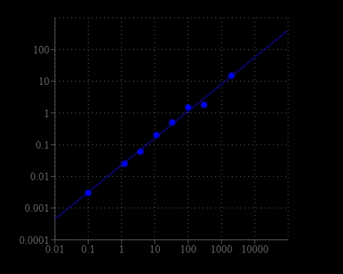
Acetylcholinesterase dose response was measured in a white/clear bottom 96-well plate with Amplite® Colorimetric Acetylcholinesterase Assay Kit using a SpectraMax microplate reader (Molecular devices). As low as 0.1 mU/well of Acetylcholinesterase can be detected with 30 minutes incubation(n=3).
Ca<sup>2+</sup> response of reticulocytes to LPA stimulation.(A) A representative image of a new methylene blue staining of RBCs from a BALB/c mouse after induction of reticulocytosis. The coloured regions depict reticulocytes analysed in (B) and (C). The arrowheads point to lysed RBCs. (B) Image of Fluo-4 loaded live RBCs. The coloured regions are transferred from (A). The dashed grey circle labels a responding RBC. (C) Intensity traces for Ca<sup>2+</sup> content of the cells marked in (B) stimulated with LPA. (D) Statistics of the maximal response of reticulocytes and the entire RBCs population without reticulocytes (referred to as erythrocytes) under control conditions and for 5 &micro;M LPA stimulation. The numbers below the boxes give the cell numbers taken from three mice. (E) AChE activity in reticulocytes and erythrocytes with and without stimulation with 5 &micro;M LPA for 15 min. The measurements comprise of a colorimetric assay based on 2&times;106 cells per measurement and the data is the average of 5 mice. *Acetylcholinesterase (AChE) activity of 2&times;106 RBCs of each population were performed using a colorimetric AChE assay kit (Amplite, AAT Bioquest, USA) following the manufacturers instructions. Source: Graph from <strong>Morphologically Homogeneous Red Blood Cells Present a Heterogeneous Response to Hormonal Stimulation</strong> by Jue Wang et al., <em>PLOS</em>, Jun. 2013.
Hippocampal levels of the AChE, CREB and neurotrophic proteins. AChE activity (A) in the hippocampus was measured with an ELISA kit. The western blotting assay for the phosphorylated CREB, BDNF, and NGF levels in hippocampal tissues (B) and BDNF levels in HT22 cell (C), and their relative intensities (D) were shown. The data are expressed as the means &plusmn; SD (n = 7 or 3). #P &lt; 0.05, ##P &lt; 0.01, and ###P &lt; 0.001 compared with the vehicle group; *P &lt; 0.05, **P &lt; 0.01, and ***P &lt; 0.001 compared with the control group. *The acetylcholinesterase (AChE) activity in the hippocampus was determined using an AChE activity assay kit (AAT Bioquest; Sunnyvale, CA, USA) according to the manufacturer&rsquo;s protocol. The absorbance at 410 nm was measured using a UV spectrophotometer. Source: Graph from <strong>Gongjin-Dan Enhances Hippocampal Memory in a Mouse Model of Scopolamine-Induced Amnesia</strong> by Jin-Seok Lee et al., <em>PLOS</em>, Aug. 2016.&nbsp;
AChE activity, mAChR1, phosphorylated CREB, and BDNF protein and gene expression as possible mechanisms of PNE action. (A) AChE activity in the hippocampus (n = 7). (B) Phosphorylated CREB and BDNF levels in the hippocampus determined by Western blotting (n = 7). (C) Quantification of phosphorylated CREB/CREB intensity. (D) Quantification of BDNF/&beta;-actin intensity. (E) Alterations in the expression of CREB1, mAChR1, BDNF, CBP and iNOS determined by real time-PCR (n = 6). Gene expression was normalized to that of &beta;-actin. Data are expressed as means &plusmn; SD. #P &lt; 0.05, ##P &lt; 0.01, ###P &lt; 0.001, compared with the na&iuml;ve group; *P &lt; 0.05, **P &lt; 0.01, ***P &lt; 0.001 compared with the control group. PNE; pine needle extract, THA; tacrine. Source: <strong>Hippocampal memory enhancing activity of pine needle extract against scopolamine-induced amnesia in a mouse model </strong>by Lee et al., <em>Scientific Reports</em>, May 2015.
The 70% ethanol extract of Spirulina maxima (SM70EE) ameliorates learning and memory impairments by inhibiting the amyloid-β (Aβ) accumulation induced by intracerebroventricular injection of Aβ1–42 in mice. Mouse hippocampal lysates were subjected to AChE assay to investigate AChE activity (n = 4 per group). Results were analyzed by one-way analysis of variance and Duncan’s multiple range test. Source: <b>Spirulina maxima Extract Ameliorates Learning and Memory Impairments via Inhibiting GSK-3β Phosphorylation Induced by Intracerebroventricular Injection of Amyloid-β 1–42 in Mice</b> by Koh et.al., <em>Int. J. Mol. Sci.</em> Nov. 2017.